Hussain Syed A, Palmer Daniel H, Syn Wing-Kin, Sacco Joseph J, Greensmith Richard M D, Elmetwali Taha, Aachi Vijay, Lloyd Bryony H, Jithesh Puthen V, Arrand John, Barton Darren, Ansari Jawaher, Sibson D Ross, James Nicholas D
Department of Molecular and Clinical Cancer Medicine, University of Liverpool, Liverpool L69 3GA, UK.
Regeneration and Repair Group, The Institute of Hepatology, Foundation of Liver Research, London SE5 9NT, UK.
Int J Oncol. 2017 Apr;50(4):1147-1159. doi: 10.3892/ijo.2017.3893. Epub 2017 Mar 2.
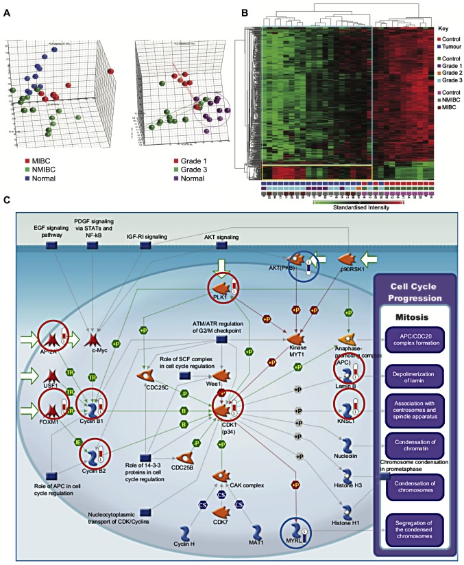
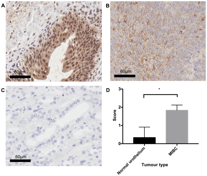
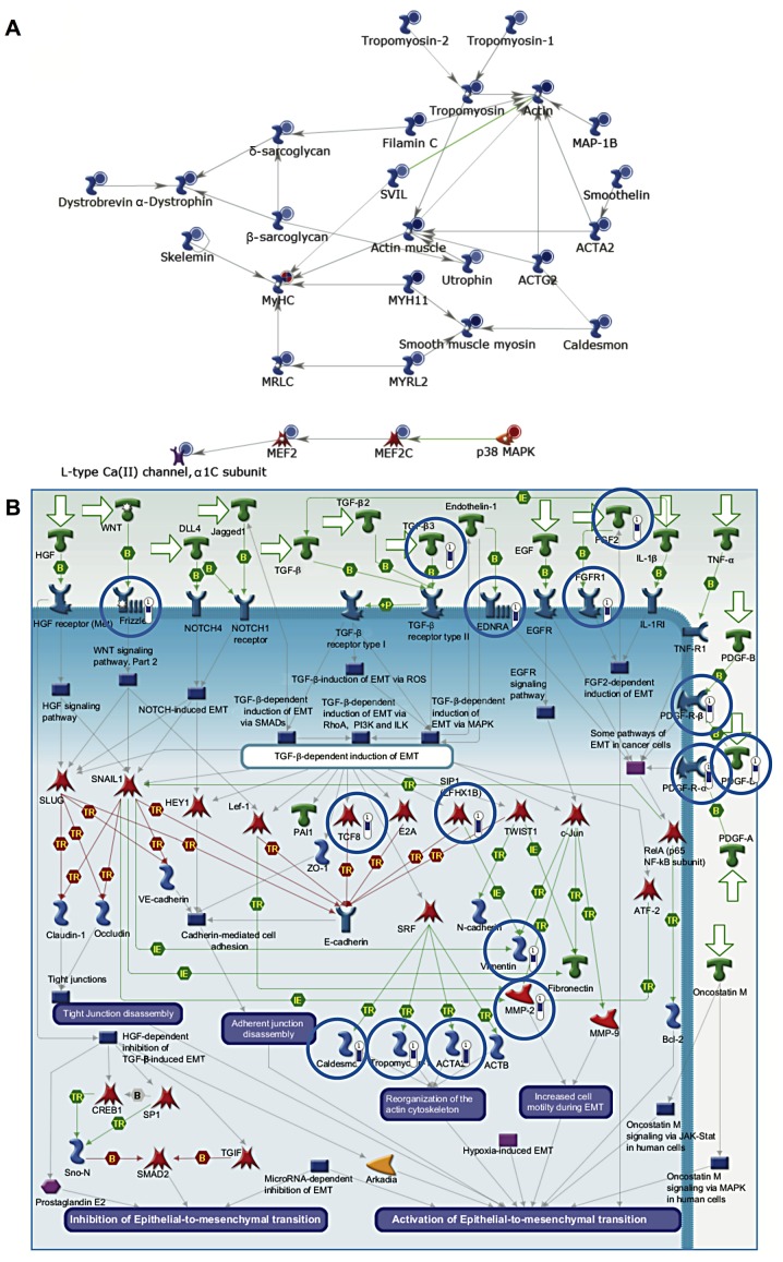
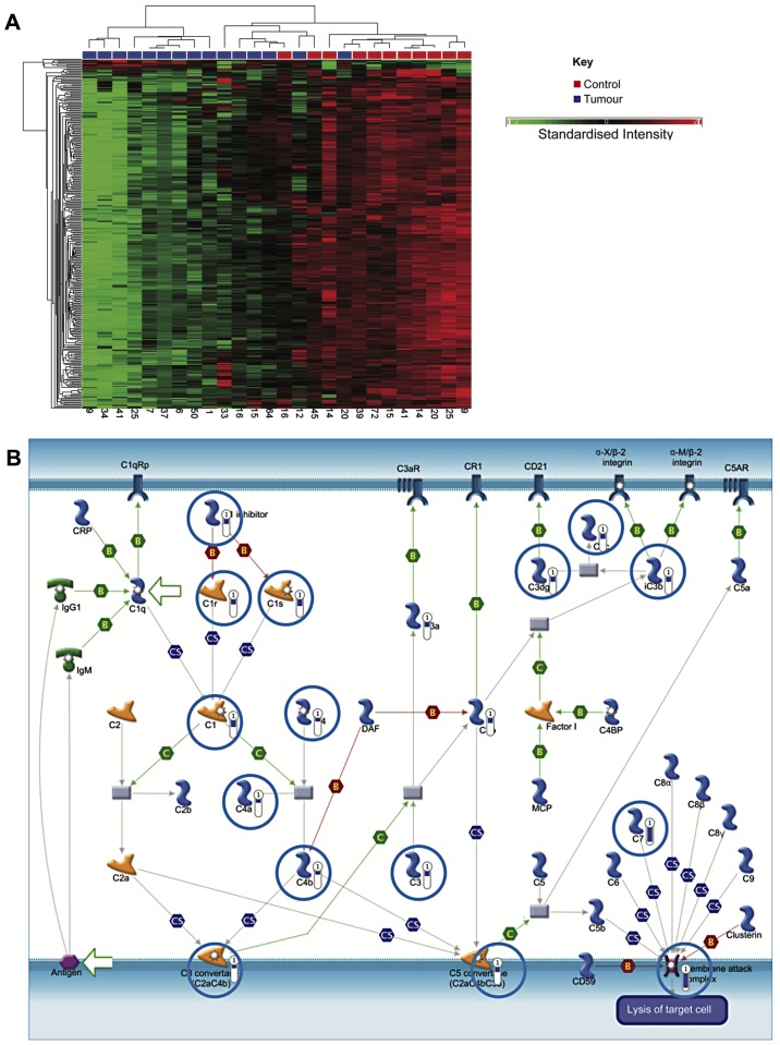
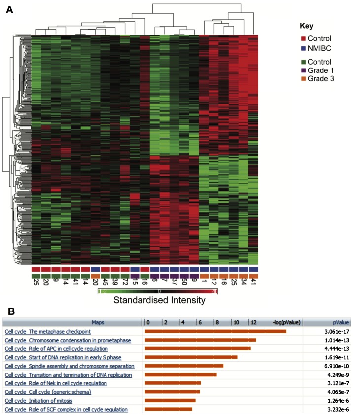
Despite advances in management, bladder cancer remains a major cause of cancer related complications. Characterisation of gene expression patterns in bladder cancer allows the identification of pathways involved in its pathogenesis, and may stimulate the development of novel therapies targeting these pathways. Between 2004 and 2005, cystoscopic bladder biopsies were obtained from 19 patients and 11 controls. These were subjected to whole transcript-based microarray analysis. Unsupervised hierarchical clustering was used to identify samples with similar expression profiles. Hypergeometric analysis was used to identify canonical pathways and curated networks having statistically significant enrichment of differentially expressed genes. Osteopontin (OPN) expression was validated by immunohistochemistry. Hierarchical clustering defined signatures, which differentiated between cancer and healthy tissue, muscle-invasive or non-muscle invasive cancer and healthy tissue, grade 1 and grade 3. Pathways associated with cell cycle and proliferation were markedly upregulated in muscle-invasive and grade 3 cancers. Genes associated with the classical complement pathway were downregulated in non-muscle invasive cancer. Osteopontin was markedly overexpressed in invasive cancer compared to healthy tissue. The present study contributes to a growing body of work on gene expression signatures in bladder cancer. The data support an important role for osteopontin in bladder cancer, and identify several pathways worthy of further investigation.
尽管在治疗方面取得了进展,但膀胱癌仍然是癌症相关并发症的主要原因。对膀胱癌基因表达模式的表征有助于识别其发病机制中涉及的途径,并可能推动针对这些途径的新型疗法的开发。在2004年至2005年期间,从19例患者和11名对照中获取了膀胱镜下膀胱活检组织。对这些组织进行了基于全转录本的微阵列分析。使用无监督层次聚类来识别具有相似表达谱的样本。使用超几何分析来识别具有统计学显著差异表达基因富集的经典途径和精选网络。通过免疫组织化学验证骨桥蛋白(OPN)的表达。层次聚类定义了特征,可区分癌症组织与健康组织、肌层浸润性或非肌层浸润性癌症与健康组织、1级和3级癌症。与细胞周期和增殖相关的途径在肌层浸润性和3级癌症中明显上调。与经典补体途径相关的基因在非肌层浸润性癌症中下调。与健康组织相比,骨桥蛋白在浸润性癌症中明显过表达。本研究为膀胱癌基因表达特征的不断积累的工作做出了贡献。数据支持骨桥蛋白在膀胱癌中的重要作用,并确定了几个值得进一步研究的途径。