Novak Marko, Perhavec Andraz, Maturen Katherine E, Pavlovic Djokic Snezana, Jereb Simona, Erzen Darja
Department of Surgical Oncology, Institute of Oncology Ljubljana, Ljubljana, Slovenia.
Department of Radiology, University of Michigan Hospitals, Ann Arbor, Michigan, USA.
Radiol Oncol. 2016 Dec 23;51(1):56-64. doi: 10.1515/raon-2016-0051. eCollection 2017 Mar 1.
Leiomyosarcoma is a rare malignant mesenchymal tumour. Some cases of leiomyosarcoma of the renal vein (LRV) have been reported in the literature, but no analysis of data and search for prognostic factors have been done so far. The aim of this review was to describe the LRV, to analyse overall survival (OS), local recurrence free survival (LRFS) and distant metastases free survival (DMFS) in LRV world case series and to identify significant predictors of OS, LRFS and DMFS.
Cases from the literature based on PubMed search and a case from our institution were included.
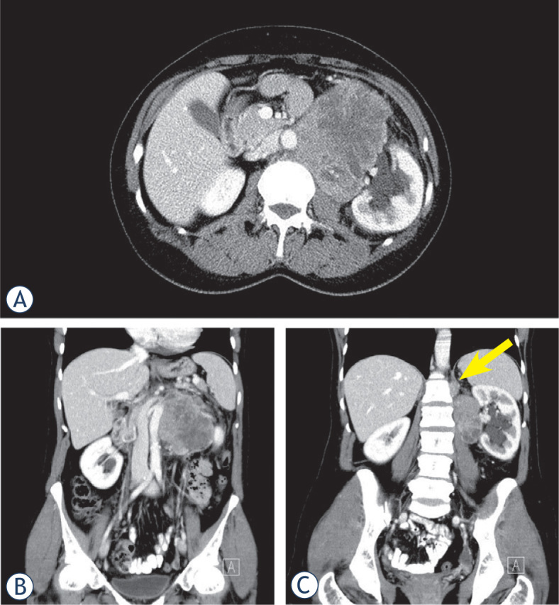
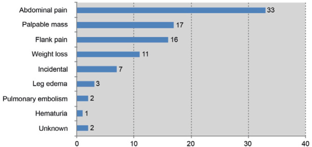
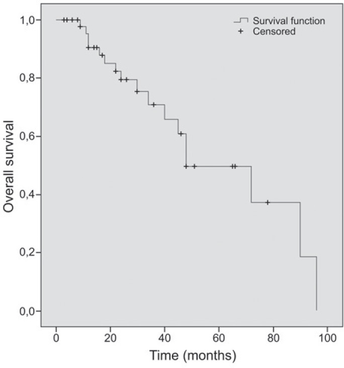
Sixty-seven patients with a mean age of 56.6 years were identified; 76.1% were women. Mean tumour size was 8.9 cm; in 68.7% located on the left side. Tumour thrombus extended into the inferior vena cava lumen in 13.4%. All patients but one underwent surgery (98.5%). After a median follow up of 24 months, the OS was 79.5%. LRFS was 83.5% after a median follow up of 21.5 months and DMFS was 76.1% after a median follow up of 22 months. Factors predictive of OS in univariate analysis were surgical margins, while factors predictive of LRFS were inferior vena cava luminal extension and grade. No factors predictive of DMFS were identified. In multivariate analysis none of the factors were predictive of OS, LRFS and DMFS.
Based on the literature review and presented case some conclusions can be made. LRV is usually located in the hilum of the kidney. It should be considered in differential diagnosis of renal and retroperitoneal masses, particularly in women over the age 40, on the left side and in the absence of haematuria. Core needle biopsy should be performed. Patients should be managed by sarcoma multidisciplinary team. LRV should be surgically removed, with negative margins.
平滑肌肉瘤是一种罕见的恶性间叶组织肿瘤。文献中已报道了一些肾静脉平滑肌肉瘤(LRV)病例,但目前尚未对数据进行分析及寻找预后因素。本综述的目的是描述LRV,分析LRV全球病例系列中的总生存期(OS)、无局部复发生存期(LRFS)和无远处转移生存期(DMFS),并确定OS、LRFS和DMFS的显著预测因素。
纳入基于PubMed搜索的文献病例及本机构的1例病例。
共确定67例患者,平均年龄56.6岁;76.1%为女性。平均肿瘤大小为8.9 cm;68.7%位于左侧。13.4%的肿瘤血栓延伸至下腔静脉腔内。除1例患者外,所有患者均接受了手术(98.5%)。中位随访24个月后,OS为79.5%。中位随访21.5个月后,LRFS为83.5%,中位随访22个月后,DMFS为76.1%。单因素分析中,预测OS的因素为手术切缘,预测LRFS的因素为下腔静脉腔内延伸和分级。未发现预测DMFS的因素。多因素分析中,无因素可预测OS、LRFS和DMFS。
基于文献综述和所呈现的病例可得出一些结论。LRV通常位于肾门。在肾及腹膜后肿块的鉴别诊断中应考虑该病,尤其是40岁以上、左侧且无血尿的女性。应进行粗针穿刺活检。患者应由肉瘤多学科团队管理。LRV应手术切除,切缘阴性。