Poombal Fnu, Ahsan Muhammad, Noor Rida, Nasir Saira, Khan Anam
Pathology, Nishtar Medical University, Multan, PAK.
Histopathology, Chughtai Institute of Pathology, Lahore, PAK.
Cureus. 2023 Sep 18;15(9):e45476. doi: 10.7759/cureus.45476. eCollection 2023 Sep.
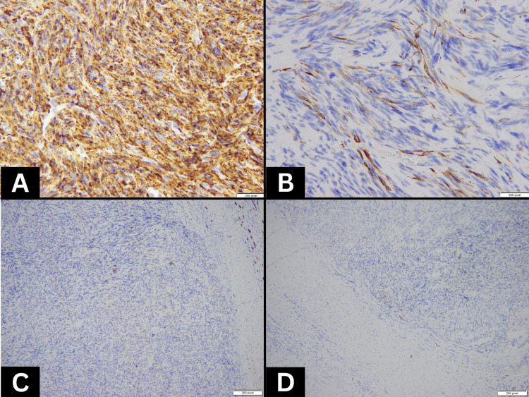
Primary leiomyosarcoma is a rare malignant kidney tumor. The diagnosis of this disease is usually made on the basis of histological examination because it lacks specific clinical or radiological characteristics. Differentiation between leiomyosarcoma and sarcomatoid renal cell carcinoma can be challenging because spindle cell morphology is observed in both tumors. Therefore, caution should be exercised when making a diagnosis of primary renal leiomyosarcoma. Both renal sarcoma and sarcomatoid renal cell carcinoma have a worse prognosis, and nephrectomy is the treatment of choice in locally resectable tumors. An example of such a tumor is discussed in relation to its diagnostic challenges. We report a case of a 35-year-old female who presented with a left renal mass. A left radical nephrectomy was performed, and a firm, tan-white, lobulated tumor (14x8x7.5 cm) was present on gross examination. A histological diagnosis of high-grade leiomyosarcoma was made on the basis of histology, positivity for caldesmon and desmin, and negative cytokeratin immunostaining. Sarcomatoid renal cell carcinoma was ruled out based on morphological findings after extensive sampling of the tumor along with negativity for CK, CD-10, and carbonic anhydrase IX immunostaining.
原发性平滑肌肉瘤是一种罕见的肾脏恶性肿瘤。由于缺乏特异性的临床或影像学特征,该疾病通常通过组织学检查来诊断。平滑肌肉瘤与肉瘤样肾细胞癌的鉴别可能具有挑战性,因为两种肿瘤均可见梭形细胞形态。因此,在诊断原发性肾平滑肌肉瘤时应谨慎。肾肉瘤和肉瘤样肾细胞癌的预后均较差,对于局部可切除的肿瘤,肾切除术是首选的治疗方法。本文结合其诊断挑战讨论了这样一个肿瘤的病例。我们报告一例35岁女性,她因左肾肿物就诊。实施了左肾根治性切除术,大体检查发现一个质地硬、呈棕白色、分叶状的肿瘤(14×8×7.5cm)。基于组织学、钙调蛋白和结蛋白阳性以及细胞角蛋白免疫染色阴性,做出了高级别平滑肌肉瘤的组织学诊断。在对肿瘤进行广泛取材以及CK、CD-10和碳酸酐酶IX免疫染色均为阴性后,基于形态学表现排除了肉瘤样肾细胞癌。