• 文献检索
  • 文档翻译
  • 深度研究
  • 学术资讯
  • Suppr Zotero 插件Zotero 插件
  • 邀请有礼
  • 套餐&价格
  • 历史记录
应用&插件
Suppr Zotero 插件Zotero 插件浏览器插件Mac 客户端Windows 客户端微信小程序
定价
高级版会员购买积分包购买API积分包
服务
文献检索文档翻译深度研究API 文档MCP 服务
关于我们
关于 Suppr公司介绍联系我们用户协议隐私条款
关注我们

Suppr 超能文献

核心技术专利:CN118964589B侵权必究
粤ICP备2023148730 号-1Suppr @ 2026

文献检索

告别复杂PubMed语法,用中文像聊天一样搜索,搜遍4000万医学文献。AI智能推荐,让科研检索更轻松。

立即免费搜索

文件翻译

保留排版,准确专业,支持PDF/Word/PPT等文件格式,支持 12+语言互译。

免费翻译文档

深度研究

AI帮你快速写综述,25分钟生成高质量综述,智能提取关键信息,辅助科研写作。

立即免费体验

实验性脑型疟疾病理学的定量脑图谱。

A quantitative brain map of experimental cerebral malaria pathology.

作者信息

Strangward Patrick, Haley Michael J, Shaw Tovah N, Schwartz Jean-Marc, Greig Rachel, Mironov Aleksandr, de Souza J Brian, Cruickshank Sheena M, Craig Alister G, Milner Danny A, Allan Stuart M, Couper Kevin N

机构信息

Faculty of Biology, Medicine and Health, University of Manchester, Manchester, United Kingdom.

Immunology Unit, Department of Infectious and Tropical Diseases, London School of Hygiene and Tropical Medicine, London, United Kingdom.

出版信息

PLoS Pathog. 2017 Mar 8;13(3):e1006267. doi: 10.1371/journal.ppat.1006267. eCollection 2017 Mar.

DOI:10.1371/journal.ppat.1006267
PMID:28273147
原文链接:https://pmc.ncbi.nlm.nih.gov/articles/PMC5358898/
Abstract

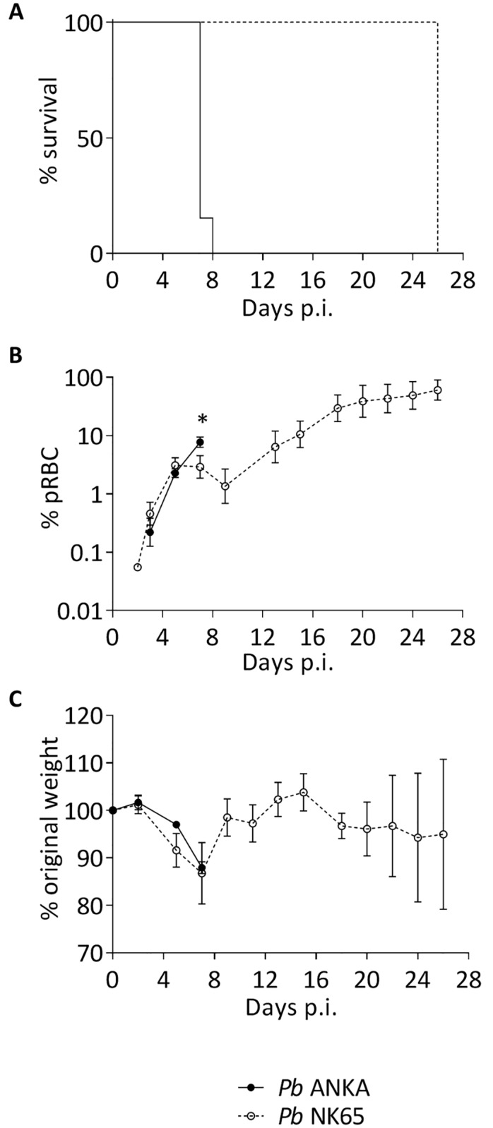
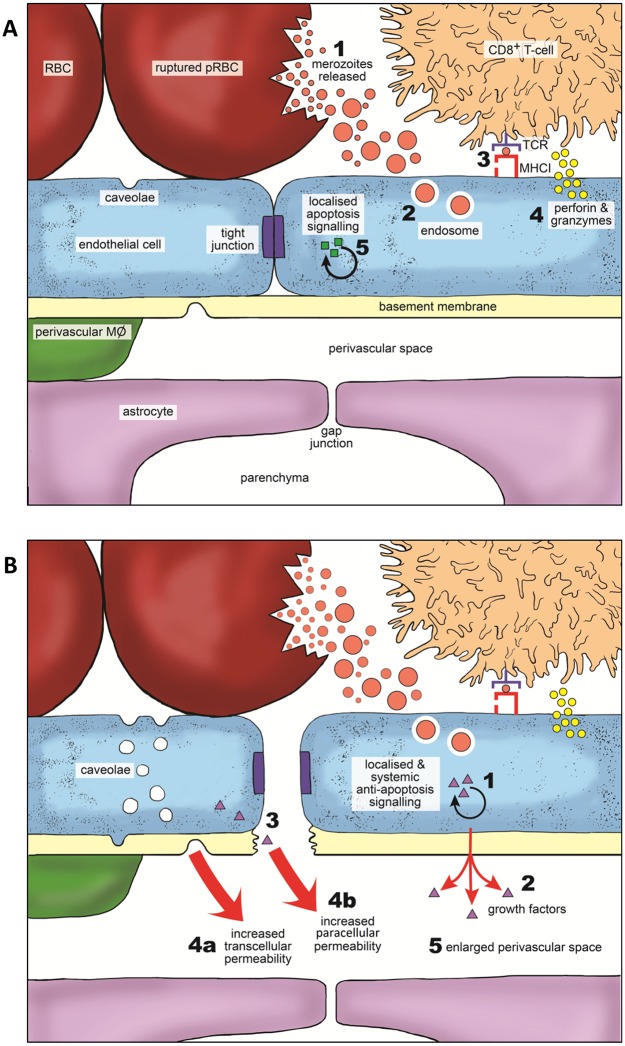
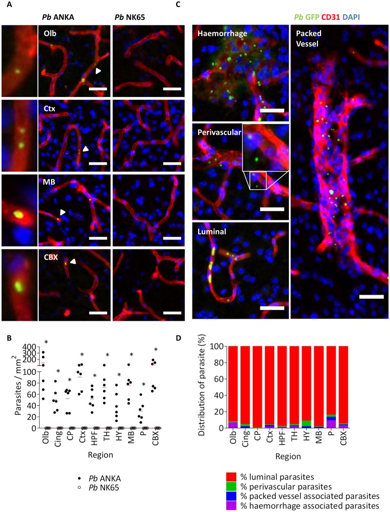
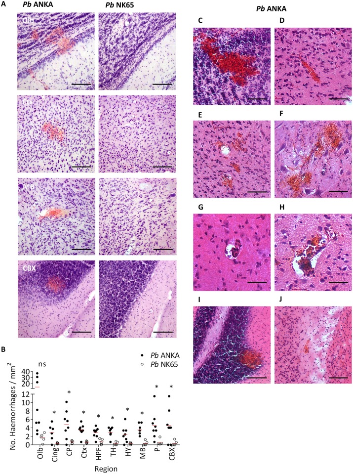
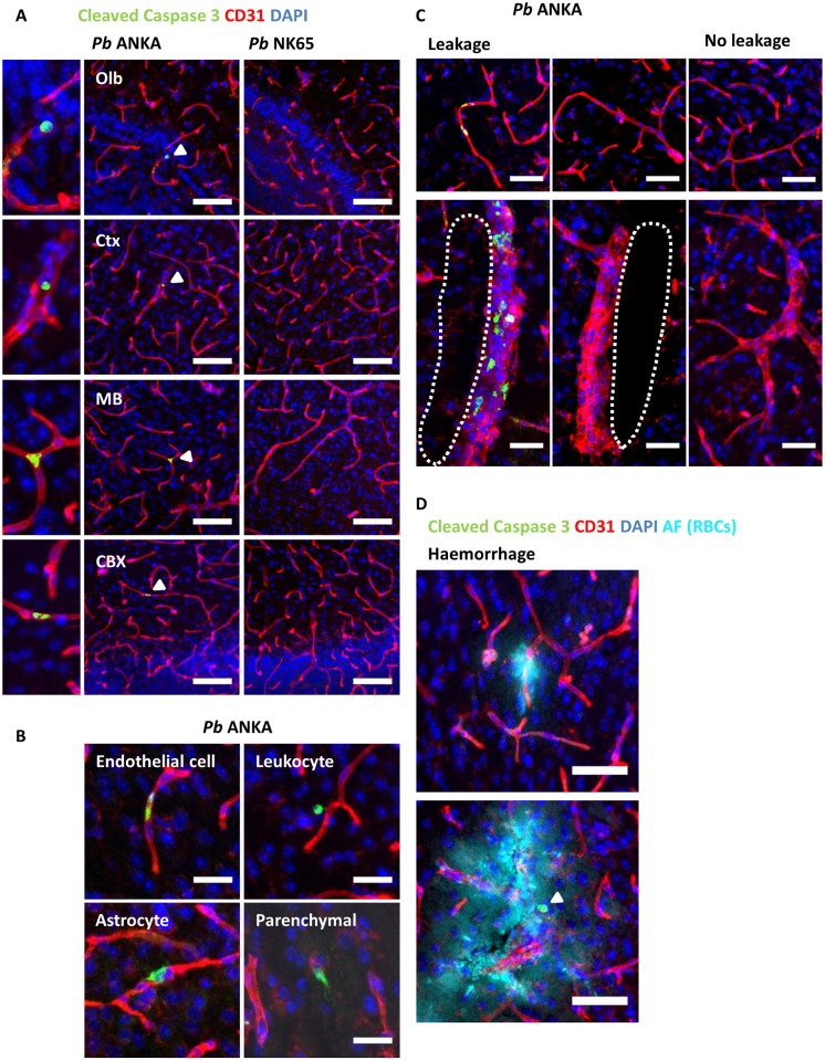
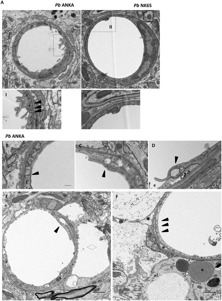
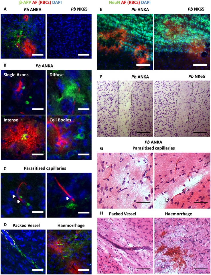
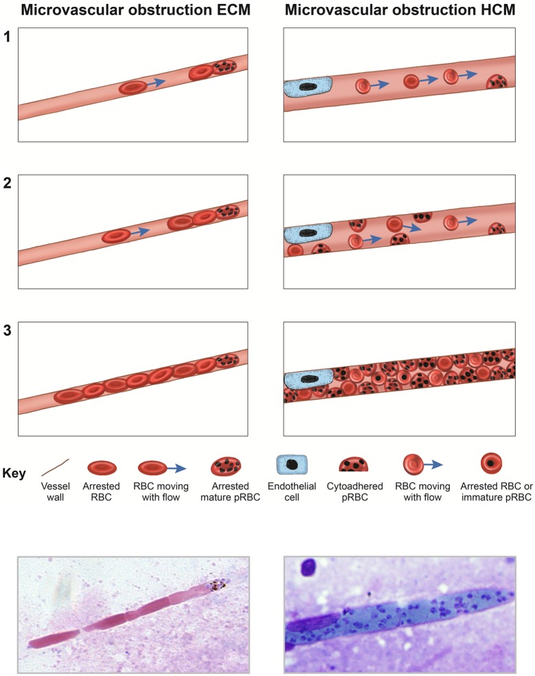
The murine model of experimental cerebral malaria (ECM) has been utilised extensively in recent years to study the pathogenesis of human cerebral malaria (HCM). However, it has been proposed that the aetiologies of ECM and HCM are distinct, and, consequently, no useful mechanistic insights into the pathogenesis of HCM can be obtained from studying the ECM model. Therefore, in order to determine the similarities and differences in the pathology of ECM and HCM, we have performed the first spatial and quantitative histopathological assessment of the ECM syndrome. We demonstrate that the accumulation of parasitised red blood cells (pRBCs) in brain capillaries is a specific feature of ECM that is not observed during mild murine malaria infections. Critically, we show that individual pRBCs appear to occlude murine brain capillaries during ECM. As pRBC-mediated congestion of brain microvessels is a hallmark of HCM, this suggests that the impact of parasite accumulation on cerebral blood flow may ultimately be similar in mice and humans during ECM and HCM, respectively. Additionally, we demonstrate that cerebrovascular CD8+ T-cells appear to co-localise with accumulated pRBCs, an event that corresponds with development of widespread vascular leakage. As in HCM, we show that vascular leakage is not dependent on extensive vascular destruction. Instead, we show that vascular leakage is associated with alterations in transcellular and paracellular transport mechanisms. Finally, as in HCM, we observed axonal injury and demyelination in ECM adjacent to diverse vasculopathies. Collectively, our data therefore shows that, despite very different presentation, and apparently distinct mechanisms, of parasite accumulation, there appear to be a number of comparable features of cerebral pathology in mice and in humans during ECM and HCM, respectively. Thus, when used appropriately, the ECM model may be useful for studying specific pathological features of HCM.

摘要

近年来,实验性脑型疟疾(ECM)的小鼠模型被广泛用于研究人类脑型疟疾(HCM)的发病机制。然而,有人提出ECM和HCM的病因不同,因此,通过研究ECM模型无法获得关于HCM发病机制的有用的机制性见解。因此,为了确定ECM和HCM病理学的异同,我们对ECM综合征进行了首次空间和定量组织病理学评估。我们证明,脑毛细血管中被寄生红细胞(pRBCs)的积累是ECM的一个特定特征,在轻度小鼠疟疾感染期间未观察到。至关重要的是,我们表明在ECM期间单个pRBCs似乎会阻塞小鼠脑毛细血管。由于pRBC介导的脑微血管充血是HCM的一个标志,这表明在ECM和HCM期间,寄生虫积累对脑血流的影响在小鼠和人类中最终可能相似。此外,我们证明脑血管CD8 + T细胞似乎与积累的pRBCs共定位,这一事件与广泛的血管渗漏的发展相对应。与HCM一样,我们表明血管渗漏不依赖于广泛的血管破坏。相反,我们表明血管渗漏与跨细胞和细胞旁转运机制的改变有关。最后,与HCM一样,我们在与各种血管病变相邻的ECM中观察到轴突损伤和脱髓鞘。因此,我们的数据总体表明,尽管寄生虫积累的表现非常不同且机制明显不同,但在ECM和HCM期间,小鼠和人类的脑病理学似乎有许多可比特征。因此,当适当使用时,ECM模型可能有助于研究HCM的特定病理特征。

https://cdn.ncbi.nlm.nih.gov/pmc/blobs/4d6e/5358898/49051d7ce463/ppat.1006267.g012.jpg
https://cdn.ncbi.nlm.nih.gov/pmc/blobs/4d6e/5358898/4c8221e97225/ppat.1006267.g001.jpg
https://cdn.ncbi.nlm.nih.gov/pmc/blobs/4d6e/5358898/1d24869f4b34/ppat.1006267.g002.jpg
https://cdn.ncbi.nlm.nih.gov/pmc/blobs/4d6e/5358898/38ec96a4d1d5/ppat.1006267.g003.jpg
https://cdn.ncbi.nlm.nih.gov/pmc/blobs/4d6e/5358898/5556398c6ed1/ppat.1006267.g004.jpg
https://cdn.ncbi.nlm.nih.gov/pmc/blobs/4d6e/5358898/8c290639e1e1/ppat.1006267.g005.jpg
https://cdn.ncbi.nlm.nih.gov/pmc/blobs/4d6e/5358898/4b18e9df68f6/ppat.1006267.g006.jpg
https://cdn.ncbi.nlm.nih.gov/pmc/blobs/4d6e/5358898/f5345733f28a/ppat.1006267.g007.jpg
https://cdn.ncbi.nlm.nih.gov/pmc/blobs/4d6e/5358898/0c6844dad3ff/ppat.1006267.g008.jpg
https://cdn.ncbi.nlm.nih.gov/pmc/blobs/4d6e/5358898/060091497d4c/ppat.1006267.g009.jpg
https://cdn.ncbi.nlm.nih.gov/pmc/blobs/4d6e/5358898/27789dbad758/ppat.1006267.g010.jpg
https://cdn.ncbi.nlm.nih.gov/pmc/blobs/4d6e/5358898/88f609d7536b/ppat.1006267.g011.jpg
https://cdn.ncbi.nlm.nih.gov/pmc/blobs/4d6e/5358898/49051d7ce463/ppat.1006267.g012.jpg

相似文献

1
A quantitative brain map of experimental cerebral malaria pathology.实验性脑型疟疾病理学的定量脑图谱。
PLoS Pathog. 2017 Mar 8;13(3):e1006267. doi: 10.1371/journal.ppat.1006267. eCollection 2017 Mar.
2
Comparison of CD8 T Cell Accumulation in the Brain During Human and Murine Cerebral Malaria.人脑和鼠脑疟疾中 CD8 T 细胞在脑内聚集的比较。
Front Immunol. 2019 Jul 24;10:1747. doi: 10.3389/fimmu.2019.01747. eCollection 2019.
3
Simultaneously targeting inflammatory response and parasite sequestration in brain to treat Experimental Cerebral Malaria.同时针对脑部炎症反应和寄生虫滞留以治疗实验性脑型疟疾。
Sci Rep. 2015 Jul 31;5:12671. doi: 10.1038/srep12671.
4
Neuroimmunological blood brain barrier opening in experimental cerebral malaria.实验性脑疟疾中的神经免疫性血脑屏障开放。
PLoS Pathog. 2012;8(10):e1002982. doi: 10.1371/journal.ppat.1002982. Epub 2012 Oct 25.
5
Young Sprague Dawley rats infected by Plasmodium berghei: A relevant experimental model to study cerebral malaria.感染伯氏疟原虫的幼年斯普拉格-道利大鼠:一种用于研究脑型疟疾的相关实验模型。
PLoS One. 2017 Jul 24;12(7):e0181300. doi: 10.1371/journal.pone.0181300. eCollection 2017.
6
Brain microvessel cross-presentation is a hallmark of experimental cerebral malaria.脑微血管交叉呈递是实验性脑疟疾的一个标志。
EMBO Mol Med. 2013 Jul;5(7):984-99. doi: 10.1002/emmm.201202273. Epub 2013 May 16.
7
CD8+ T Cells Induce Fatal Brainstem Pathology during Cerebral Malaria via Luminal Antigen-Specific Engagement of Brain Vasculature.CD8 + T细胞通过脑微血管腔面抗原特异性结合在脑型疟疾期间诱发致命性脑干病理改变。
PLoS Pathog. 2016 Dec 1;12(12):e1006022. doi: 10.1371/journal.ppat.1006022. eCollection 2016 Dec.
8
Critical role of IL-33 receptor ST2 in experimental cerebral malaria development.白细胞介素-33受体ST2在实验性脑型疟疾发展中的关键作用。
Eur J Immunol. 2015 May;45(5):1354-65. doi: 10.1002/eji.201445206. Epub 2015 Mar 20.
9
IFN-γ-producing CD4+ T cells promote experimental cerebral malaria by modulating CD8+ T cell accumulation within the brain.IFN-γ 产生的 CD4+T 细胞通过调节大脑内 CD8+T 细胞的积累来促进实验性脑疟疾。
J Immunol. 2012 Jul 15;189(2):968-79. doi: 10.4049/jimmunol.1200688. Epub 2012 Jun 20.
10
Using a new three-dimensional CUBIC tissue-clearing method to examine the brain during experimental cerebral malaria.采用一种新的三维 CUBIC 组织通透化方法检测实验性脑疟疾期间的大脑。
Int Immunol. 2021 Oct 29;33(11):587-594. doi: 10.1093/intimm/dxab060.

引用本文的文献

1
Characterization of experimental cerebral malaria by volumetric MRI A comparative study across the sexes.通过容积磁共振成像对实验性脑型疟疾进行特征描述:一项跨性别比较研究
PLoS One. 2025 Aug 18;20(8):e0328693. doi: 10.1371/journal.pone.0328693. eCollection 2025.
2
CD8 T cells promote tubule-interstitial damage in malaria-induced acute kidney injury.CD8 T细胞在疟疾诱导的急性肾损伤中促进肾小管间质损伤。
Front Cell Infect Microbiol. 2025 Jun 30;15:1561806. doi: 10.3389/fcimb.2025.1561806. eCollection 2025.
3
Self-assembling dendrimer nanodrug formulations for decreased hERG-related toxicity and enhanced therapeutic efficacy.

本文引用的文献

1
Quantification of Cerebral Vascular Architecture using Two-photon Microscopy in a Mouse Model of HIV-induced Neuroinflammation.在HIV诱导的神经炎症小鼠模型中使用双光子显微镜对脑血管结构进行定量分析。
J Vis Exp. 2016 Jan 12(107):e53582. doi: 10.3791/53582.
2
Perivascular Arrest of CD8+ T Cells Is a Signature of Experimental Cerebral Malaria.CD8 + T细胞的血管周围停滞是实验性脑疟疾的一个特征。
PLoS Pathog. 2015 Nov 12;11(11):e1005210. doi: 10.1371/journal.ppat.1005210. eCollection 2015.
3
Activated Brain Endothelial Cells Cross-Present Malaria Antigen.
用于降低hERG相关毒性并提高治疗效果的自组装树枝状大分子纳米药物制剂。
Sci Adv. 2025 Jun 27;11(26):eadu9948. doi: 10.1126/sciadv.adu9948. Epub 2025 Jun 25.
4
Neuronal Damage in Murine Experimental Cerebral Malaria, Implications for Neuronal Repair and Sequelae.小鼠实验性脑型疟疾中的神经元损伤,对神经元修复和后遗症的影响。
Cells. 2025 May 30;14(11):807. doi: 10.3390/cells14110807.
5
Combination astragaloside IV and artesunate preserves blood-brain barrier integrity by modulating astrocytes and tight junction proteins in Plasmodium yoelii infection.黄芪甲苷IV与青蒿琥酯联合使用通过调节约氏疟原虫感染中的星形胶质细胞和紧密连接蛋白来维持血脑屏障的完整性。
Int J Parasitol Drugs Drug Resist. 2025 May 28;28:100598. doi: 10.1016/j.ijpddr.2025.100598.
6
ICAM-1/CD18-mediated sequestration of parasitized phagocytes in cortical capillaries promotes neuronal colonization by Toxoplasma gondii.ICAM-1/CD18介导的寄生吞噬细胞在皮质毛细血管中的隔离促进了弓形虫对神经元的定植。
Nat Commun. 2025 Apr 14;16(1):3529. doi: 10.1038/s41467-025-58655-z.
7
Nilotinib attenuates vascular pathology in experimental cerebral malaria.尼罗替尼减轻实验性脑型疟疾中的血管病变。
Blood Adv. 2025 May 27;9(10):2473-2488. doi: 10.1182/bloodadvances.2024015364.
8
Unveiling new perspectives about the onset of neurological and cognitive deficits in cerebral malaria: exploring cellular and neurochemical mechanisms.揭示脑型疟疾中神经和认知缺陷发病的新观点:探索细胞和神经化学机制。
Front Cell Infect Microbiol. 2025 Feb 6;15:1506282. doi: 10.3389/fcimb.2025.1506282. eCollection 2025.
9
The spatiotemporal transcriptional profiling of murine brain during cerebral malaria progression and after artemisinin treatment.小鼠脑在脑型疟疾进展过程中和青蒿素治疗后的时空转录谱分析。
Nat Commun. 2025 Feb 11;16(1):1540. doi: 10.1038/s41467-024-52223-7.
10
Non-cerebral malaria: does such a thing exist?非脑型疟疾:有这种病吗?
Mem Inst Oswaldo Cruz. 2025 Feb 3;120:e240223. doi: 10.1590/0074-02760240223. eCollection 2025.
活化的脑内皮细胞交叉提呈疟疾抗原。
PLoS Pathog. 2015 Jun 5;11(6):e1004963. doi: 10.1371/journal.ppat.1004963. eCollection 2015 Jun.
4
Microvascular obstruction and endothelial activation are independently associated with the clinical manifestations of severe falciparum malaria in adults: an observational study.微血管阻塞和内皮细胞激活与成人重症恶性疟的临床表现独立相关:一项观察性研究。
BMC Med. 2015 May 27;13:122. doi: 10.1186/s12916-015-0365-9.
5
Brain swelling and death in children with cerebral malaria.脑肿胀和死亡的儿童与脑疟疾。
N Engl J Med. 2015 Mar 19;372(12):1126-37. doi: 10.1056/NEJMoa1400116.
6
Experimental cerebral malaria pathogenesis--hemodynamics at the blood brain barrier.实验性脑型疟发病机制——血脑屏障处的血流动力学
PLoS Pathog. 2014 Dec 4;10(12):e1004528. doi: 10.1371/journal.ppat.1004528. eCollection 2014 Dec.
7
Real-time imaging reveals the dynamics of leukocyte behaviour during experimental cerebral malaria pathogenesis.实时成像揭示了实验性脑型疟疾发病过程中白细胞行为的动态变化。
PLoS Pathog. 2014 Jul 17;10(7):e1004236. doi: 10.1371/journal.ppat.1004236. eCollection 2014 Jul.
8
Stuck in a rut? Reconsidering the role of parasite sequestration in severe malaria syndromes.陷入困境?重新考虑寄生虫隔离在严重疟疾综合征中的作用。
Trends Parasitol. 2013 Dec;29(12):585-92. doi: 10.1016/j.pt.2013.10.004. Epub 2013 Nov 5.
9
Cytoadherence of Plasmodium berghei-infected red blood cells to murine brain and lung microvascular endothelial cells in vitro.体外感染伯氏疟原虫的红细胞对鼠脑和肺微血管内皮细胞的细胞黏附作用。
Infect Immun. 2013 Nov;81(11):3984-91. doi: 10.1128/IAI.00428-13. Epub 2013 Aug 12.
10
Brain microvessel cross-presentation is a hallmark of experimental cerebral malaria.脑微血管交叉呈递是实验性脑疟疾的一个标志。
EMBO Mol Med. 2013 Jul;5(7):984-99. doi: 10.1002/emmm.201202273. Epub 2013 May 16.