Chen Jiayun, Bai Yunmeng, He Xueling, Xiao Wei, Chen Lina, Wong Yin Kwan, Wang Chen, Gao Peng, Cheng Guangqing, Xu Liting, Yang Chuanbin, Liao Fulong, Han Guang, Sun Jichao, Xu Chengchao, Wang Jigang
State Key Laboratory for Quality Ensurance and Sustainable Use of Dao-di Herbs, Artemisinin Research Center, and Institute of Chinese Materia Medica, China Academy of Chinese Medical Sciences, Beijing, 100700, China.
Department of Critical Care Medicine, Guangdong Provincial Clinical Research Center for Geriatrics, Shenzhen Clinical Research Center for Geriatric, Shenzhen People's Hospital (The Second Clinical Medical College, Jinan University; The First Affiliated Hospital, Southern University of Science and Technology), Shenzhen, 518020, Guangdong, China.
Nat Commun. 2025 Feb 11;16(1):1540. doi: 10.1038/s41467-024-52223-7.
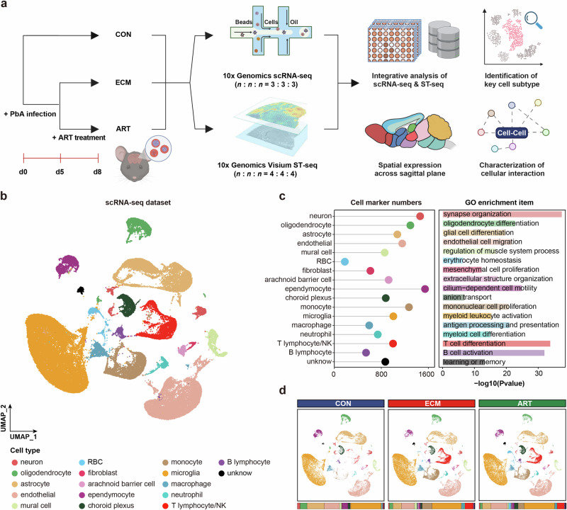
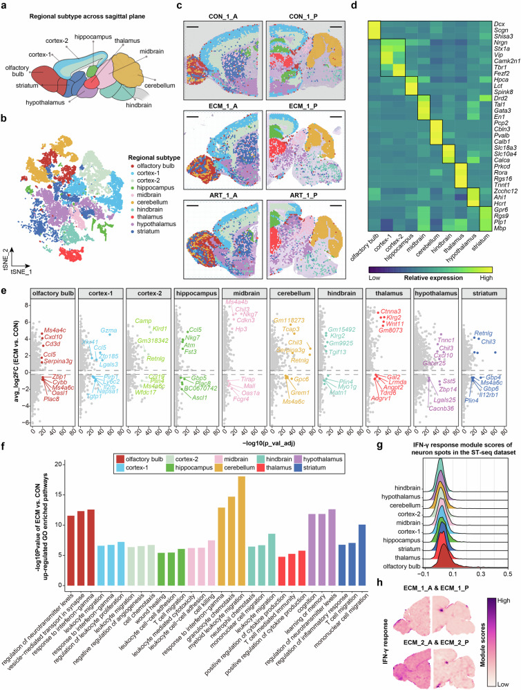
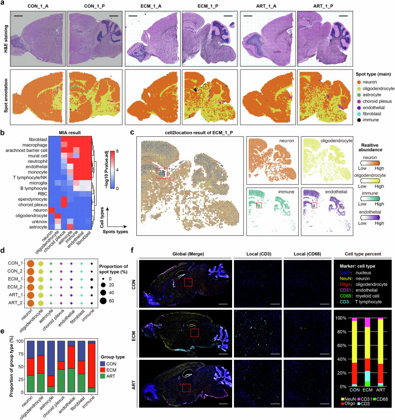
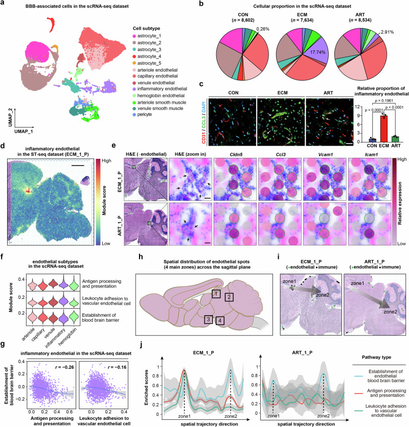
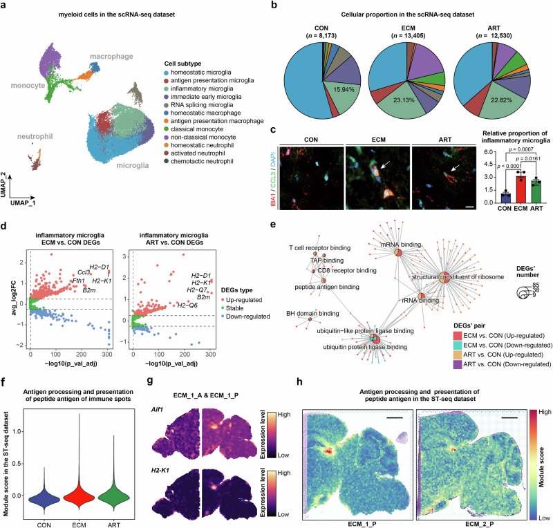
Cerebral malaria (CM) is a severe encephalopathy caused by Plasmodium parasite infection, resulting in thousands of annual deaths and neuro-cognitive sequelae even after anti-malarial drugs treatment. Despite efforts to dissect the mechanism, the cellular transcriptomic reprogramming within the spatial context remains elusive. Here, we constructed single-cell and spatial transcriptome atlases of experimental CM (ECM) male murine brain tissues with or without artesunate (ART) treatment. We identified activated inflammatory endothelial cells during ECM, characterized by a disrupted blood-brain barrier, increased antigen presentation, and leukocyte adhesion. We also observed that inflammatory microglia enhance antigen presentation pathway such as MHC-I to CD8 cytotoxic T cells. The latter underwent an inflammatory state transition with up-regulated cytokine expression and cytotoxic activity. Multi-omics analysis revealed that the activated interferon-gamma response of injured neurons during ECM and persisted after ART treatment. Overall, our research provides valuable resources for understanding malaria parasite-host interaction mechanisms and adjuvant therapy development.
脑型疟疾(CM)是由疟原虫感染引起的严重脑病,即使在使用抗疟药物治疗后,每年仍会导致数千人死亡以及神经认知后遗症。尽管人们努力剖析其机制,但在空间背景下的细胞转录组重编程仍不清楚。在此,我们构建了接受或未接受青蒿琥酯(ART)治疗的实验性脑型疟疾(ECM)雄性小鼠脑组织的单细胞和空间转录组图谱。我们在ECM期间鉴定出活化的炎性内皮细胞,其特征为血脑屏障破坏、抗原呈递增加和白细胞粘附。我们还观察到炎性小胶质细胞增强了诸如MHC-I至CD8细胞毒性T细胞的抗原呈递途径。后者经历了炎性状态转变,细胞因子表达和细胞毒性活性上调。多组学分析显示,ECM期间受损神经元的干扰素-γ反应被激活,并在ART治疗后持续存在。总体而言,我们的研究为理解疟原虫与宿主的相互作用机制以及辅助治疗的开发提供了有价值的资源。