Henkel Anne S, LeCuyer Brian, Olivares Shantel, Green Richard M
Division of Gastroenterology and Hepatology, Feinberg School of Medicine, Northwestern University, Chicago, Illinois.
Cell Mol Gastroenterol Hepatol. 2016 Dec 10;3(2):261-271. doi: 10.1016/j.jcmgh.2016.11.006. eCollection 2017 Mar.
BACKGROUND & AIMS: Cholestasis promotes endoplasmic reticulum (ER) stress in the liver, however, the effect of ER stress on hepatic bile acid metabolism is unknown. We aim to determine the effect of ER stress on hepatic bile acid synthesis and transport in mice.
ER stress was induced pharmacologically in C57BL/6J mice and human hepatoma (HepG2) cells. The hepatic expression of genes controlling bile acid synthesis and transport was determined. To measure the activity of the primary bile acid synthetic pathway, the concentration of 7α-hydroxy-4-cholesten-3-1 was measured in plasma.
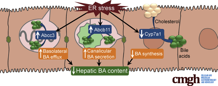
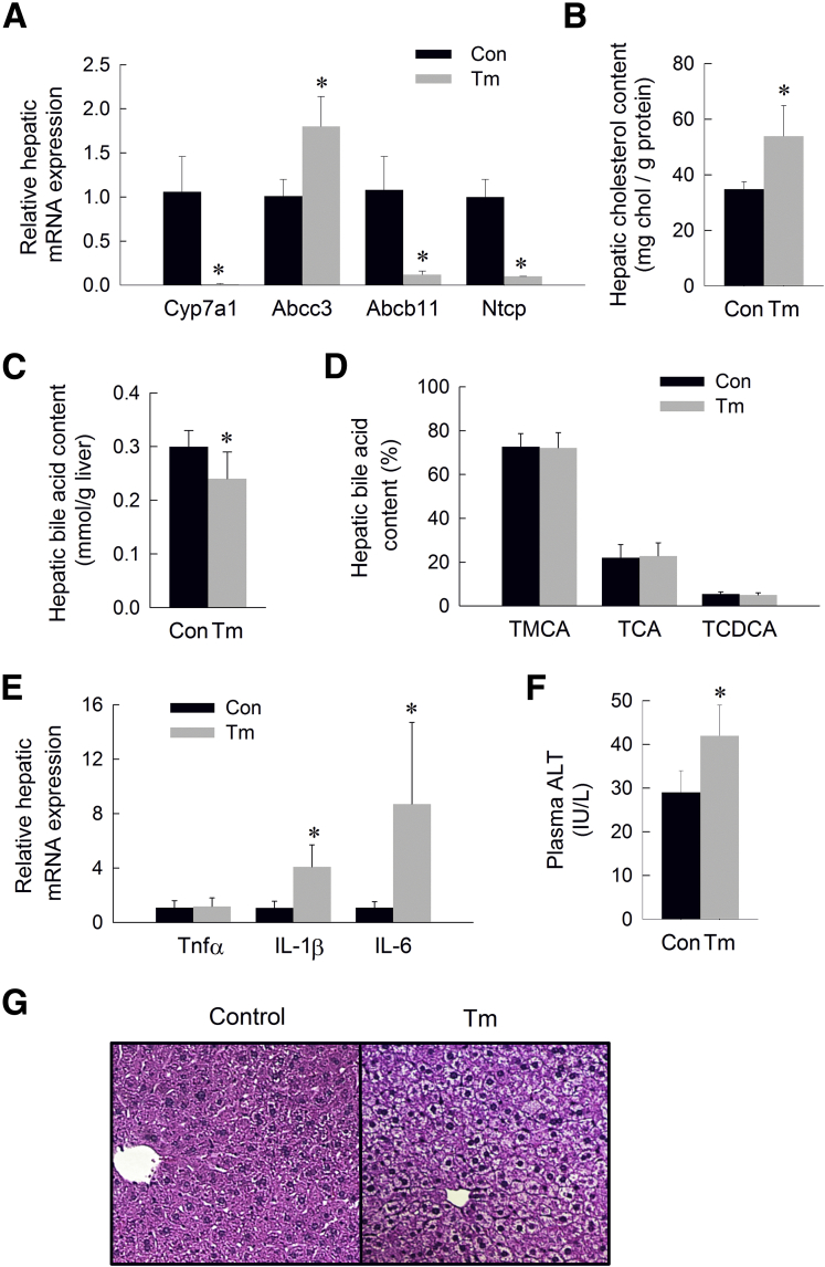
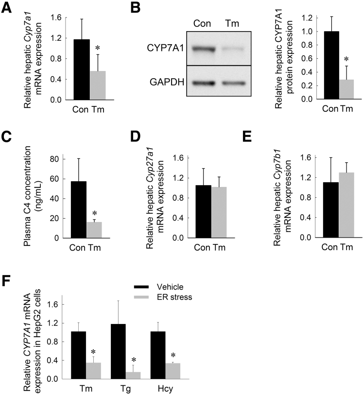
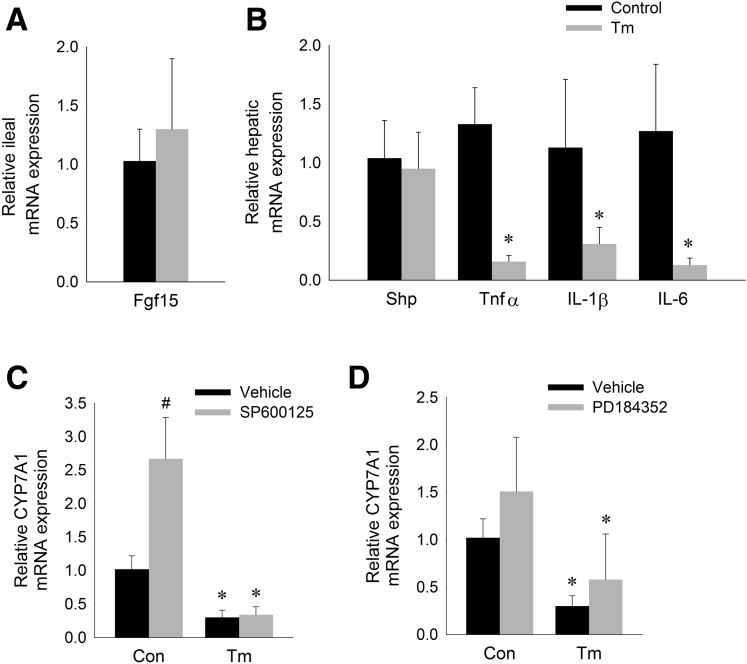
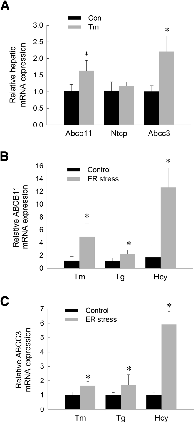
Induction of ER stress in mice and HepG2 cells rapidly suppressed the hepatic expression of the primary bile acid synthetic enzyme, cholesterol 7α-hydroxylase. Plasma levels of 7α-hydroxy-4-cholesten-3-1 were reduced in mice subjected to ER stress, indicating impaired bile acid synthesis. Induction of ER stress in mice and HepG2 cells increased expression of the bile salt export pump (adenosine triphosphate binding cassette []) and a bile salt efflux pump (). The observed regulation of , , and occurred in the absence of hepatic inflammatory cytokine activation and was not dependent on activation of hepatic small heterodimer partner or intestinal fibroblast growth factor 15. Consistent with suppressed bile acid synthesis and enhanced bile acid export from hepatocytes, prolonged ER stress decreased the hepatic bile acid content in mice.
Induction of ER stress in mice suppresses bile acid synthesis and enhances bile acid removal from hepatocytes independently of established bile acid regulatory pathways. These data show a novel function of the ER stress response in regulating bile acid metabolism.
胆汁淤积会促进肝脏内质网(ER)应激,然而,内质网应激对肝脏胆汁酸代谢的影响尚不清楚。我们旨在确定内质网应激对小鼠肝脏胆汁酸合成和转运的影响。
通过药理学方法在C57BL/6J小鼠和人肝癌(HepG2)细胞中诱导内质网应激。测定控制胆汁酸合成和转运的基因在肝脏中的表达。为了测量初级胆汁酸合成途径的活性,检测血浆中7α-羟基-4-胆甾烯-3-酮的浓度。
在小鼠和HepG2细胞中诱导内质网应激可迅速抑制初级胆汁酸合成酶胆固醇7α-羟化酶在肝脏中的表达。内质网应激小鼠的血浆7α-羟基-4-胆甾烯-3-酮水平降低,表明胆汁酸合成受损。在小鼠和HepG2细胞中诱导内质网应激可增加胆盐输出泵(三磷酸腺苷结合盒转运体)和一种胆盐外排泵的表达。观察到的对、和的调节发生在肝脏炎性细胞因子未激活的情况下,且不依赖于肝脏小异源二聚体伴侣或肠成纤维细胞生长因子15的激活。与胆汁酸合成受抑制和肝细胞胆汁酸输出增强一致,内质网应激延长会降低小鼠肝脏胆汁酸含量。
在小鼠中诱导内质网应激可抑制胆汁酸合成,并独立于已有的胆汁酸调节途径增强肝细胞对胆汁酸的清除。这些数据显示了内质网应激反应在调节胆汁酸代谢中的新功能。