Liyanage Sumedha, Dassanayake Rohan S, Bouyanfif Amal, Rajakaruna Erandathi, Ramalingam Latha, Moustaid-Moussa Naima, Abidi Noureddine
Fiber and Biopolymer Research Institute, Department of Plant and Soil Science, Texas Tech University, Lubbock, TX 79403, USA.
Fiber and Biopolymer Research Institute, Department of Plant and Soil Science, Texas Tech University, Lubbock, TX 79403, USA; Department of Nutritional Sciences, Texas Tech University, Lubbock, TX 79409, USA.
MethodsX. 2017 Feb 2;4:118-127. doi: 10.1016/j.mex.2017.01.006. eCollection 2017.
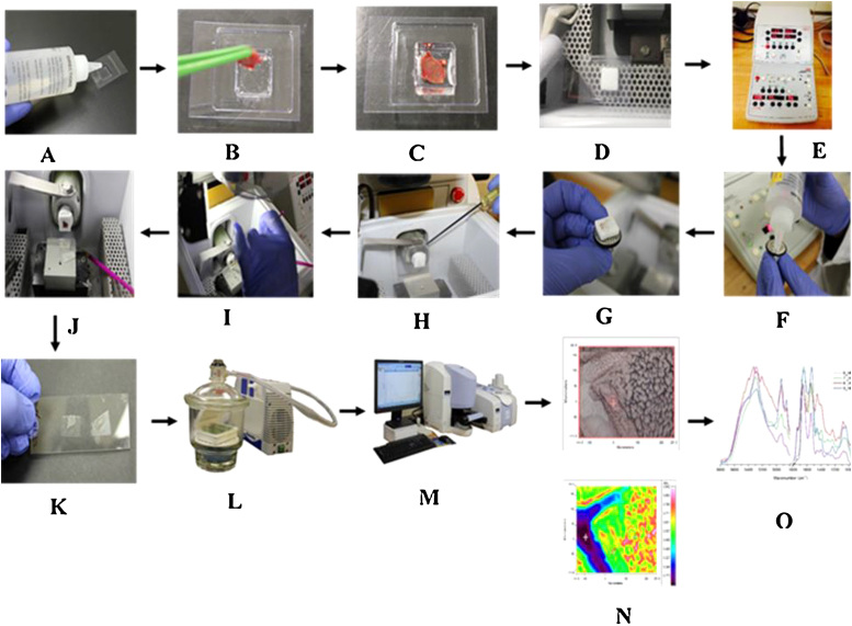
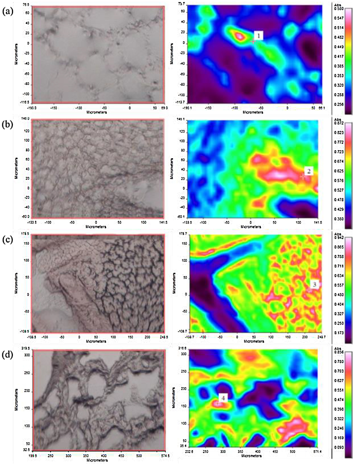
In Fourier transform infrared (FTIR) microspectrocopy, the tissue preparation method is crucial, especially how the tissue is cryo-sectioned prior to the imaging requires special consideration. Having a temperature difference between the cutting blade and the specimen holder of the cryostat greatly affects the quality of the sections. Therefore, we have developed an optimal protocol for cryo-sectioning of biological tissues by varying the temperature of both the cutting blade and the specimen holder. Using this protocol, we successfully cryo-sectioned four different difficult-to-section tissues including white adipose tissue (WAT), brown adipose tissue (BAT), lung, and liver. The optimal temperatures that required to be maintained at the cutting blade and the specimen holder for the cryo-sectioning of WAT, BAT, lung, and liver are (-25, -20 °C), (-25, -20 °C), (-17, -13 °C) and (-15, -5 °C), respectively. The optimized protocol developed in this study produced high quality cryo-sections with sample thickness of 8-10 μm, as well as high quality trans-reflectance mode FTIR microspectroscopic images for the tissue sections. •Use of cryostat technique to make thin sections of biological samples for FTIR microspectroscopy imaging.•Optimized cryostat temperature conditions by varying the temperatures at the cutting blade and specimen holder to obtain high quality sections of difficult-to-handle tissues.•FTIR imaging is used to obtain chemical information from cryo-sectioned samples with no interference of the conventional paraffin-embedding agent and chemicals.
在傅里叶变换红外(FTIR)显微光谱分析中,组织制备方法至关重要,尤其是在成像前组织如何进行冷冻切片需要特别考虑。低温恒温器的切割刀片与样品架之间存在温差会极大地影响切片质量。因此,我们通过改变切割刀片和样品架的温度,开发了一种用于生物组织冷冻切片的优化方案。使用该方案,我们成功地对四种不同的难切片组织进行了冷冻切片,包括白色脂肪组织(WAT)、棕色脂肪组织(BAT)、肺和肝脏。对WAT、BAT、肺和肝脏进行冷冻切片时,切割刀片和样品架需要保持的最佳温度分别为(-25,-20℃)、(-25,-20℃)、(-17,-13℃)和(-15,-5℃)。本研究中开发的优化方案产生了高质量的冷冻切片,样品厚度为8 - 10μm,以及用于组织切片的高质量透反射模式FTIR显微光谱图像。•使用低温恒温器技术制作生物样品的薄切片用于FTIR显微光谱成像。•通过改变切割刀片和样品架的温度优化低温恒温器温度条件,以获得难以处理的组织的高质量切片。•FTIR成像用于从冷冻切片样品中获取化学信息,不受传统石蜡包埋剂和化学物质的干扰。