• 文献检索
  • 文档翻译
  • 深度研究
  • 学术资讯
  • Suppr Zotero 插件Zotero 插件
  • 邀请有礼
  • 套餐&价格
  • 历史记录
应用&插件
Suppr Zotero 插件Zotero 插件浏览器插件Mac 客户端Windows 客户端微信小程序
定价
高级版会员购买积分包购买API积分包
服务
文献检索文档翻译深度研究API 文档MCP 服务
关于我们
关于 Suppr公司介绍联系我们用户协议隐私条款
关注我们

Suppr 超能文献

核心技术专利:CN118964589B侵权必究
粤ICP备2023148730 号-1Suppr @ 2026

文献检索

告别复杂PubMed语法,用中文像聊天一样搜索,搜遍4000万医学文献。AI智能推荐,让科研检索更轻松。

立即免费搜索

文件翻译

保留排版,准确专业,支持PDF/Word/PPT等文件格式,支持 12+语言互译。

免费翻译文档

深度研究

AI帮你快速写综述,25分钟生成高质量综述,智能提取关键信息,辅助科研写作。

立即免费体验

静态猪瓣膜间质细胞培养物的稳健生成

Robust Generation of Quiescent Porcine Valvular Interstitial Cell Cultures.

作者信息

Porras Ana M, van Engeland Nicole C A, Marchbanks Evelyn, McCormack Ann, Bouten Carlijn V C, Yacoub Magdi H, Latif Najma, Masters Kristyn S

机构信息

Department of Biomedical Engineering, University of Wisconsin-Madison, Madison, WI.

Department of Biomedical Engineering, Eindhoven University of Technology, Eindhoven, The Netherlands.

出版信息

J Am Heart Assoc. 2017 Mar 14;6(3):e005041. doi: 10.1161/JAHA.116.005041.

DOI:10.1161/JAHA.116.005041
PMID:28292746
原文链接:https://pmc.ncbi.nlm.nih.gov/articles/PMC5524027/
Abstract

BACKGROUND

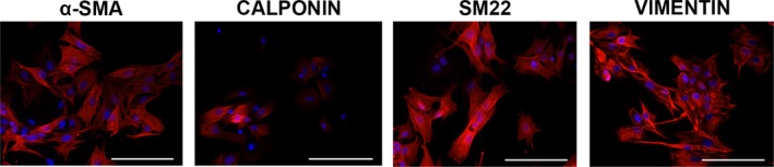
Valvular interstitial cells (VICs) in the healthy aortic valve leaflet exhibit a quiescent phenotype, with <5% of VICs exhibiting an activated phenotype. Yet, in vitro culture of VICs on tissue culture polystyrene surfaces in standard growth medium results in rapid transformation to an activated phenotype in >90% of cells. The inability to preserve a healthy VIC phenotype during in vitro studies has hampered the elucidation of mechanisms involved in calcific aortic valve disease. This study describes the generation of quiescent populations of porcine VICs in 2-dimensional in vitro culture and their utility in studying valve pathobiology.

METHODS AND RESULTS

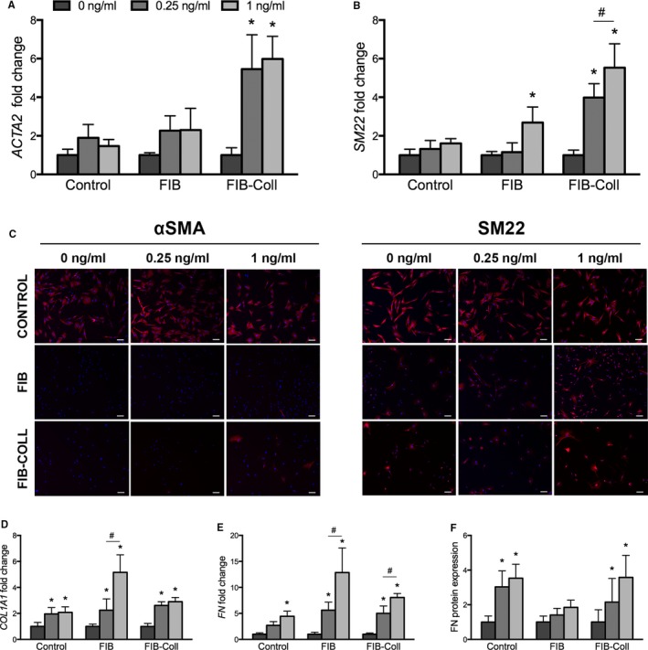
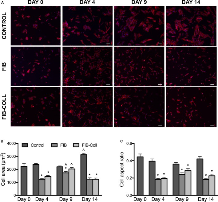
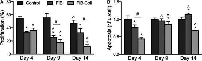
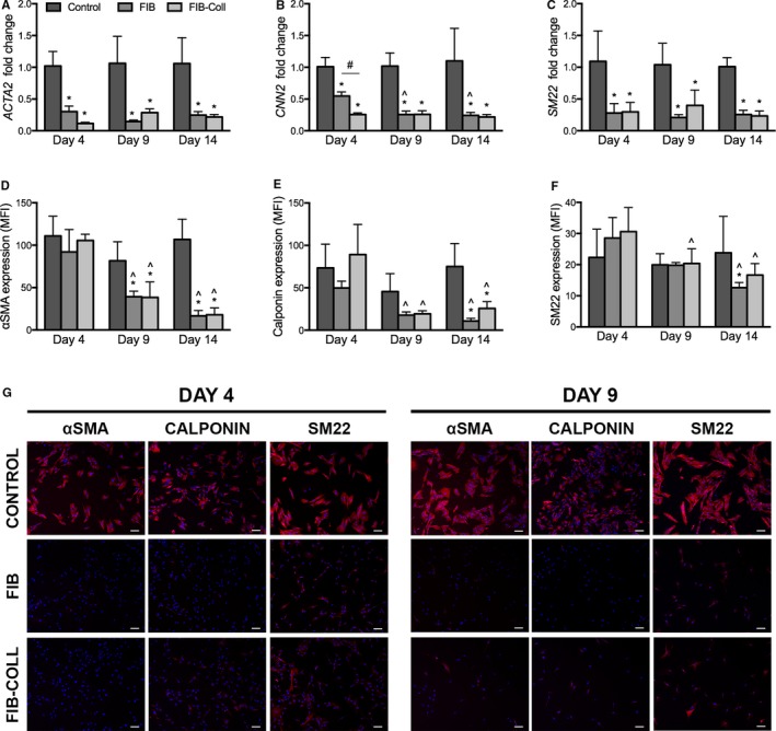
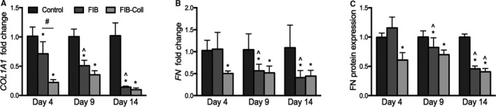
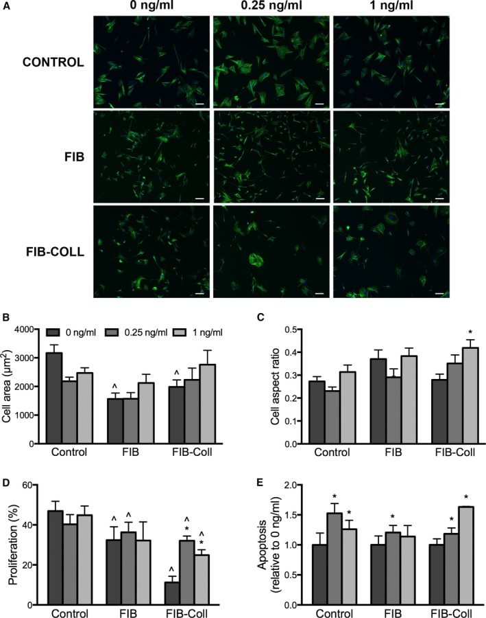
Within 4 days of isolation from fresh porcine hearts, VICs cultured in standard growth conditions were predominantly myofibroblastic (activated VICs). This myofibroblastic phenotype was partially reversed within 4 days, and fully reversed within 9 days, following application of a combination of a fibroblast media formulation with culture on collagen coatings. Specifically, culture in this combination significantly reduced several markers of VIC activation, including proliferation, apoptosis, α-smooth muscle actin expression, and matrix production, relative to standard growth conditions. Moreover, VICs raised in a fibroblast media formulation with culture on collagen coatings exhibited dramatically increased sensitivity to treatment with transforming growth factor β1, a known pathological stimulus, compared with VICs raised in either standard culture or medium with a fibroblast media formulation.

CONCLUSIONS

The approach using a fibroblast media formulation with culture on collagen coatings generates quiescent VICs that more accurately mimic a healthy VIC population and thus has the potential to transform the study of the mechanisms of VIC activation and dysfunction involved in the early stages of calcific aortic valve disease.

摘要

背景

健康主动脉瓣叶中的瓣膜间质细胞(VICs)表现出静止表型,只有不到5%的VICs表现出激活表型。然而,在标准生长培养基中,将VICs在组织培养聚苯乙烯表面进行体外培养会导致超过90%的细胞迅速转变为激活表型。在体外研究过程中无法保持健康的VIC表型,这阻碍了对钙化性主动脉瓣疾病相关机制的阐明。本研究描述了在二维体外培养中产生猪VICs静止群体的方法及其在研究瓣膜病理生物学中的应用。

方法与结果

从新鲜猪心脏分离出的VICs,在标准生长条件下培养4天内,主要为肌成纤维细胞(激活的VICs)。在应用成纤维细胞培养基配方并在胶原蛋白涂层上培养后,这种肌成纤维细胞表型在4天内部分逆转,在9天内完全逆转。具体而言,与标准生长条件相比,这种组合培养显著降低了VIC激活的几个标志物,包括增殖、凋亡、α-平滑肌肌动蛋白表达和基质产生。此外,与在标准培养或含成纤维细胞培养基配方的培养基中培养的VICs相比,在成纤维细胞培养基配方并在胶原蛋白涂层上培养的VICs对已知病理刺激物转化生长因子β1的处理表现出显著提高的敏感性。

结论

使用成纤维细胞培养基配方并在胶原蛋白涂层上培养的方法可产生更准确模拟健康VIC群体的静止VICs,因此有可能改变对钙化性主动脉瓣疾病早期阶段VIC激活和功能障碍机制的研究。

https://cdn.ncbi.nlm.nih.gov/pmc/blobs/506f/5524027/5142be2150c5/JAH3-6-e005041-g007.jpg
https://cdn.ncbi.nlm.nih.gov/pmc/blobs/506f/5524027/0f4967594741/JAH3-6-e005041-g001.jpg
https://cdn.ncbi.nlm.nih.gov/pmc/blobs/506f/5524027/a71959ce0105/JAH3-6-e005041-g002.jpg
https://cdn.ncbi.nlm.nih.gov/pmc/blobs/506f/5524027/b50a7f2b432f/JAH3-6-e005041-g003.jpg
https://cdn.ncbi.nlm.nih.gov/pmc/blobs/506f/5524027/812915eb6534/JAH3-6-e005041-g004.jpg
https://cdn.ncbi.nlm.nih.gov/pmc/blobs/506f/5524027/6a1112e6f15d/JAH3-6-e005041-g005.jpg
https://cdn.ncbi.nlm.nih.gov/pmc/blobs/506f/5524027/f316a3e158c4/JAH3-6-e005041-g006.jpg
https://cdn.ncbi.nlm.nih.gov/pmc/blobs/506f/5524027/5142be2150c5/JAH3-6-e005041-g007.jpg

相似文献

1
Robust Generation of Quiescent Porcine Valvular Interstitial Cell Cultures.静态猪瓣膜间质细胞培养物的稳健生成
J Am Heart Assoc. 2017 Mar 14;6(3):e005041. doi: 10.1161/JAHA.116.005041.
2
Transforming growth factor-β1 promotes fibrosis but attenuates calcification of valvular tissue applied as a three-dimensional calcific aortic valve disease model.转化生长因子-β1 促进纤维化,但可减轻作为三维钙化主动脉瓣疾病模型的瓣膜组织的钙化。
Am J Physiol Heart Circ Physiol. 2020 Nov 1;319(5):H1123-H1141. doi: 10.1152/ajpheart.00651.2019. Epub 2020 Sep 28.
3
Simulation of early calcific aortic valve disease in a 3D platform: A role for myofibroblast differentiation.3D平台中早期钙化性主动脉瓣疾病的模拟:肌成纤维细胞分化的作用。
J Mol Cell Cardiol. 2016 May;94:13-20. doi: 10.1016/j.yjmcc.2016.03.004. Epub 2016 Mar 17.
4
Secreted Factors From Proinflammatory Macrophages Promote an Osteoblast-Like Phenotype in Valvular Interstitial Cells.促炎巨噬细胞分泌的因子促进瓣膜间质细胞向成骨细胞样表型转化。
Arterioscler Thromb Vasc Biol. 2020 Nov;40(11):e296-e308. doi: 10.1161/ATVBAHA.120.315261. Epub 2020 Sep 17.
5
Valvular interstitial cells suppress calcification of valvular endothelial cells.瓣膜间质细胞抑制瓣膜内皮细胞的钙化。
Atherosclerosis. 2015 Sep;242(1):251-260. doi: 10.1016/j.atherosclerosis.2015.07.008. Epub 2015 Jul 17.
6
Interstitial cells in calcified aortic valves have reduced differentiation potential and stem cell-like properties.钙化主动脉瓣间质细胞分化潜能降低,具有干细胞样特性。
Sci Rep. 2019 Sep 10;9(1):12934. doi: 10.1038/s41598-019-49016-0.
7
Activated p300 acetyltransferase activity modulates aortic valvular calcification with osteogenic transdifferentiation and downregulation of Klotho.活化的p300乙酰转移酶活性通过成骨转分化和Klotho下调来调节主动脉瓣钙化。
Int J Cardiol. 2017 Apr 1;232:271-279. doi: 10.1016/j.ijcard.2017.01.005. Epub 2017 Jan 5.
8
ADAMTS5 Deficiency in Calcified Aortic Valves Is Associated With Elevated Pro-Osteogenic Activity in Valvular Interstitial Cells.钙化主动脉瓣中ADAMTS5缺乏与瓣膜间质细胞中促骨生成活性升高有关。
Arterioscler Thromb Vasc Biol. 2017 Jul;37(7):1339-1351. doi: 10.1161/ATVBAHA.117.309021. Epub 2017 May 25.
9
Sex-related differences in matrix remodeling and early osteogenic markers in aortic valvular interstitial cells.主动脉瓣间质细胞中基质重塑和早期成骨标志物的性别差异。
Heart Vessels. 2017 Feb;32(2):217-228. doi: 10.1007/s00380-016-0909-8. Epub 2016 Oct 19.
10
Manipulation of valve composition to elucidate the role of collagen in aortic valve calcification.通过调控瓣膜成分阐明胶原蛋白在主动脉瓣钙化中的作用。
BMC Cardiovasc Disord. 2014 Mar 1;14:29. doi: 10.1186/1471-2261-14-29.

引用本文的文献

1
Extracellular Matrix Dynamics in Aortic Valve Health and Disease: Insights into Fibrocalcific Remodeling and Creation of Biomimetic Platforms.主动脉瓣健康与疾病中的细胞外基质动力学:对纤维钙化重塑及仿生平台构建的见解
J Heart Valve Soc. 2024 Oct-Dec;1(1). doi: 10.1177/30494826241296675. Epub 2024 Dec 26.
2
In vitro model assesses the susceptibility of polymeric scaffolds for material-driven heart valve regeneration to calcification.体外模型评估了用于材料驱动心脏瓣膜再生的聚合物支架对钙化的敏感性。
In Vitro Model. 2025 Jul 15;4(2):157-175. doi: 10.1007/s44164-025-00090-x. eCollection 2025 Aug.
3
Mechanical Modulation, Physiological Roles, and Imaging Innovations of Intercellular Calcium Waves in Living Systems.

本文引用的文献

1
Calcific Aortic Valve Disease Is Associated with Layer-Specific Alterations in Collagen Architecture.钙化性主动脉瓣疾病与胶原蛋白结构的层特异性改变有关。
PLoS One. 2016 Sep 29;11(9):e0163858. doi: 10.1371/journal.pone.0163858. eCollection 2016.
2
Active tissue stiffness modulation controls valve interstitial cell phenotype and osteogenic potential in 3D culture.活性组织硬度调节在三维培养中控制瓣膜间质细胞表型和成骨潜能。
Acta Biomater. 2016 May;36:42-54. doi: 10.1016/j.actbio.2016.03.007. Epub 2016 Mar 3.
3
Cells and extracellular matrix interplay in cardiac valve disease: because age matters.
活体细胞间钙波的机械调制、生理作用及成像创新
Cancers (Basel). 2025 May 31;17(11):1851. doi: 10.3390/cancers17111851.
4
Determining sex differences in aortic valve myofibroblast responses to drug combinations identified using a digital medicine platform.利用数字医学平台确定主动脉瓣成肌纤维细胞对联合用药反应中的性别差异。
Sci Adv. 2025 Jun 6;11(23):eadu2695. doi: 10.1126/sciadv.adu2695.
5
Trymethylamine-N-oxide, a gut-derived metabolite, induces myofibroblastic activation of valvular interstitial cells through endoplasmic reticulum stress.氧化三甲胺是一种源自肠道的代谢产物,它通过内质网应激诱导瓣膜间质细胞发生肌成纤维细胞活化。
bioRxiv. 2025 Feb 11:2025.02.06.636980. doi: 10.1101/2025.02.06.636980.
6
A three-dimensional valve-on-chip microphysiological system implicates cell cycle progression, cholesterol metabolism and protein homeostasis in early calcific aortic valve disease progression.一种三维片上瓣膜微生理系统揭示了早期钙化性主动脉瓣疾病进展过程中的细胞周期进程、胆固醇代谢和蛋白质稳态。
Acta Biomater. 2024 Sep 15;186:167-184. doi: 10.1016/j.actbio.2024.07.036. Epub 2024 Jul 30.
7
Aortic valve cell microenvironment: Considerations for developing a valve-on-chip.主动脉瓣细胞微环境:开发芯片上瓣膜的考量因素。
Biophys Rev (Melville). 2021 Dec 10;2(4):041303. doi: 10.1063/5.0063608. eCollection 2021 Dec.
8
Inhibition of valve mesenchymal stromal cell calcium deposition by bFGF through alternative polyadenylation regulation of the CAT gene.bFGF 通过调控 CAT 基因可变聚腺苷酸化抑制心脏瓣膜间质细胞钙沉积。
BMC Cardiovasc Disord. 2024 Feb 28;24(1):128. doi: 10.1186/s12872-024-03775-5.
9
Human and porcine aortic valve endothelial and interstitial cell isolation and characterization.人和猪主动脉瓣内皮细胞与间质细胞的分离及鉴定。
Front Cardiovasc Med. 2023 Jun 20;10:1151028. doi: 10.3389/fcvm.2023.1151028. eCollection 2023.
10
Identifying molecular and functional similarities and differences between human primary cardiac valve interstitial cells and ventricular fibroblasts.识别人类原发性心脏瓣膜间质细胞与心室成纤维细胞之间的分子和功能异同。
Front Bioeng Biotechnol. 2023 Mar 27;11:1102487. doi: 10.3389/fbioe.2023.1102487. eCollection 2023.
细胞和细胞外基质在心脏瓣膜病中的相互作用:因为年龄很重要。
Basic Res Cardiol. 2016 Mar;111(2):16. doi: 10.1007/s00395-016-0534-9. Epub 2016 Feb 1.
4
Development of Aortic Valve Disease in Familial Hypercholesterolemic Swine: Implications for Elucidating Disease Etiology.家族性高胆固醇血症猪主动脉瓣疾病的发展:对阐明疾病病因的意义。
J Am Heart Assoc. 2015 Oct 27;4(10):e002254. doi: 10.1161/JAHA.115.002254.
5
Microarray analyses to quantify advantages of 2D and 3D hydrogel culture systems in maintaining the native valvular interstitial cell phenotype.微阵列分析以量化二维和三维水凝胶培养系统在维持天然瓣膜间质细胞表型方面的优势。
Biomaterials. 2016 Jan;74:31-41. doi: 10.1016/j.biomaterials.2015.09.035. Epub 2015 Sep 28.
6
Modulation of human valve interstitial cell phenotype and function using a fibroblast growth factor 2 formulation.使用成纤维细胞生长因子2制剂调节人瓣膜间质细胞表型和功能。
PLoS One. 2015 Jun 4;10(6):e0127844. doi: 10.1371/journal.pone.0127844. eCollection 2015.
7
Cardiac valve cells and their microenvironment--insights from in vitro studies.心脏瓣膜细胞及其微环境——来自体外研究的见解
Nat Rev Cardiol. 2014 Dec;11(12):715-27. doi: 10.1038/nrcardio.2014.162. Epub 2014 Oct 14.
8
In vitro models of aortic valve calcification: solidifying a system.主动脉瓣钙化的体外模型:建立一个系统
Cardiovasc Pathol. 2015 Jan-Feb;24(1):1-10. doi: 10.1016/j.carpath.2014.08.003. Epub 2014 Aug 15.
9
Directing valvular interstitial cell myofibroblast-like differentiation in a hybrid hydrogel platform.在混合水凝胶平台中引导瓣膜间质细胞向肌成纤维细胞样分化。
Adv Healthc Mater. 2015 Jan 7;4(1):121-30. doi: 10.1002/adhm.201400029. Epub 2014 Jun 24.
10
Modelling mammalian cellular quiescence.模拟哺乳动物细胞静止状态。
Interface Focus. 2014 Jun 6;4(3):20130074. doi: 10.1098/rsfs.2013.0074.