Boufelli Gabriela, Giannotti Marcelo A, Ruiz Carlos A, Barros Nestor de, Chala Luciano F, Maesaka Jonathan Y, Goncalves Rodrigo, Bresciani Bárbara H, Vianna Paula, Soares José M, Baracat Edmund C, Filassi José R
Department of Gynecology and Obstetrics.
Department of Pathology, Hospital das Clínicas, Faculdade de Medicina, Universidade de São Paulo, São Paulo, Brazil.
Eur J Cancer Prev. 2018 Jul;27(4):310-314. doi: 10.1097/CEJ.0000000000000343.
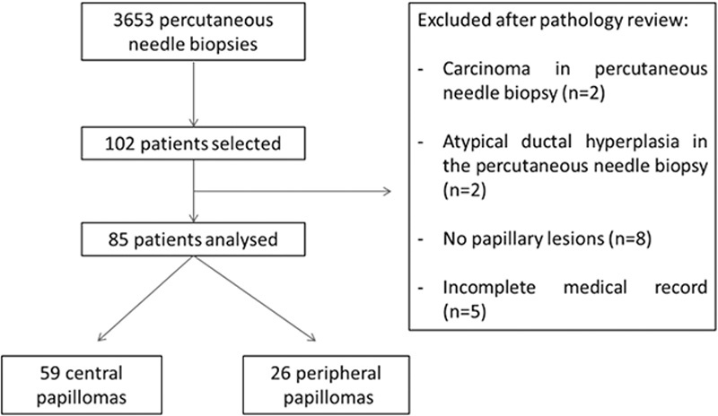
The distinction between benign and malignant papilloma of the breast through percutaneous needle biopsy can be difficult because of limited samples; the underestimation rate can be up to 25%. The aim of this study is to identify clinical and histological factors associated with underestimation, invasive ductal carcinoma, or ductal in-situ carcinoma (DCIS) of the breast found in surgical specimens from papillary lesions. This may contribute toward selection of patients for a follow-up strategy without the need for surgical excision. From a database of 3563 patients, we identified 85 with intraductal papilloma between 2007 and 2013 who had undergone breast-imaging studies, percutaneous needle biopsy, and surgical resection of the lesion. Central papillomas normally present with a palpable mass, whereas peripheral papillomas generally do not have clinical manifestations (microcalcifications); both central and peripheral papillomas were related to atypical lesions, 13.5 and 15.4%, respectively. Among the 59 cases of central papillomas, there were four cases of underestimation, three DCIS and one invasive ductal carcinoma (6.8%). Among the 26 cases of peripheral papillomas, there was one case of DCIS (3.8%), with a total underestimation rate of 5.8%; all underestimated lesions measured more than 1 cm. The median size was 11 mm at mammography and 19 mm at ultrasound. Our data suggest that lesions less than 1 cm in size, without atypia and concordant imaging and clinical findings, may not require surgical resection.
由于样本有限,通过经皮穿刺活检来区分乳腺良性和恶性乳头状瘤可能存在困难;低估率可达25%。本研究的目的是确定与乳头状病变手术标本中发现的乳腺低估、浸润性导管癌或导管原位癌(DCIS)相关的临床和组织学因素。这可能有助于选择无需手术切除即可进行后续随访策略的患者。在一个包含3563例患者的数据库中,我们确定了2007年至2013年间85例患有导管内乳头状瘤且接受过乳腺影像学检查、经皮穿刺活检及病变手术切除的患者。中央型乳头状瘤通常表现为可触及的肿块,而周围型乳头状瘤一般无临床表现(微钙化);中央型和周围型乳头状瘤均与非典型病变相关,分别为13.5%和15.4%。在59例中央型乳头状瘤病例中,有4例被低估,3例为DCIS,1例为浸润性导管癌(6.8%)。在26例周围型乳头状瘤病例中,有1例为DCIS(3.8%),总低估率为5.8%;所有被低估的病变均大于1厘米。乳腺钼靶检查时病变的中位大小为11毫米,超声检查时为19毫米。我们的数据表明,大小小于1厘米、无非典型性且影像学和临床检查结果一致的病变可能无需手术切除。