• 文献检索
  • 文档翻译
  • 深度研究
  • 学术资讯
  • Suppr Zotero 插件Zotero 插件
  • 邀请有礼
  • 套餐&价格
  • 历史记录
应用&插件
Suppr Zotero 插件Zotero 插件浏览器插件Mac 客户端Windows 客户端微信小程序
定价
高级版会员购买积分包购买API积分包
服务
文献检索文档翻译深度研究API 文档MCP 服务
关于我们
关于 Suppr公司介绍联系我们用户协议隐私条款
关注我们

Suppr 超能文献

核心技术专利:CN118964589B侵权必究
粤ICP备2023148730 号-1Suppr @ 2026

文献检索

告别复杂PubMed语法,用中文像聊天一样搜索,搜遍4000万医学文献。AI智能推荐,让科研检索更轻松。

立即免费搜索

文件翻译

保留排版,准确专业,支持PDF/Word/PPT等文件格式,支持 12+语言互译。

免费翻译文档

深度研究

AI帮你快速写综述,25分钟生成高质量综述,智能提取关键信息,辅助科研写作。

立即免费体验

乳腺孤立性扩张导管的筛查:图文综述

Screening a Solitary Dilated Duct in the Breast: A Pictorial Essay.

作者信息

Ayres Veronica Jorge, Ramalho Luciana Costa, Wludarski Sheila Cristina Lordelo, Fleury Eduardo de Faria Castro

机构信息

Department of Radiology of Instituto Brasileiro do Controle do Câncer, São Paulo, SP, 03102-002, Brazil.

Department of Pathology of Instituto Hermes Pardini SA, Belo Horizonte, MG, Brazil.

出版信息

Breast Cancer (Dove Med Press). 2021 Aug 12;13:505-512. doi: 10.2147/BCTT.S307842. eCollection 2021.

DOI:10.2147/BCTT.S307842
PMID:34413680
原文链接:https://pmc.ncbi.nlm.nih.gov/articles/PMC8369045/
Abstract

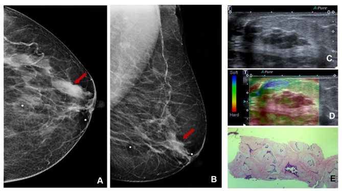
A solitary dilated duct visualized by mammography is a rare event. According to the latest edition of BI-RADS it is classified as category 4. This series of cases shows complementary ultrasound of a solitary dilated duct can reduce false-positive results on mammography.

摘要

乳腺钼靶检查显示的孤立性扩张导管是一种罕见情况。根据最新版的乳腺影像报告和数据系统(BI-RADS),它被归类为4类。这一系列病例表明,对孤立性扩张导管进行超声检查可以减少乳腺钼靶检查的假阳性结果。

https://cdn.ncbi.nlm.nih.gov/pmc/blobs/ecef/8369045/2d02b4f4e0aa/BCTT-13-505-g0008.jpg
https://cdn.ncbi.nlm.nih.gov/pmc/blobs/ecef/8369045/f497c349a182/BCTT-13-505-g0001.jpg
https://cdn.ncbi.nlm.nih.gov/pmc/blobs/ecef/8369045/5863402fa33a/BCTT-13-505-g0002.jpg
https://cdn.ncbi.nlm.nih.gov/pmc/blobs/ecef/8369045/f0232d97e8e3/BCTT-13-505-g0003.jpg
https://cdn.ncbi.nlm.nih.gov/pmc/blobs/ecef/8369045/20abca0ad659/BCTT-13-505-g0004.jpg
https://cdn.ncbi.nlm.nih.gov/pmc/blobs/ecef/8369045/eb50b1634f9c/BCTT-13-505-g0005.jpg
https://cdn.ncbi.nlm.nih.gov/pmc/blobs/ecef/8369045/f801a0a54738/BCTT-13-505-g0006.jpg
https://cdn.ncbi.nlm.nih.gov/pmc/blobs/ecef/8369045/35a09dc51cd7/BCTT-13-505-g0007.jpg
https://cdn.ncbi.nlm.nih.gov/pmc/blobs/ecef/8369045/2d02b4f4e0aa/BCTT-13-505-g0008.jpg

相似文献

1
Screening a Solitary Dilated Duct in the Breast: A Pictorial Essay.乳腺孤立性扩张导管的筛查:图文综述
Breast Cancer (Dove Med Press). 2021 Aug 12;13:505-512. doi: 10.2147/BCTT.S307842. eCollection 2021.
2
Solitary dilated duct identified at mammography: outcomes analysis.乳腺 X 线摄影检查中发现的孤立性扩张导管:结局分析。
AJR Am J Roentgenol. 2010 Feb;194(2):378-82. doi: 10.2214/AJR.09.2944.
3
Assessment of BI-RADS category 4 lesions detected with screening mammography and screening US: utility of MR imaging.乳腺 X 线筛查和超声筛查检出 BI-RADS 4 类病变的评估:磁共振成像的应用。
Radiology. 2015 Feb;274(2):343-51. doi: 10.1148/radiol.14140645. Epub 2014 Sep 29.
4
[Ultrasound examination of breast microcalcifications: luxury or necessity?].[乳腺微钙化的超声检查:是奢侈品还是必需品?]
J Radiol. 2006 Dec;87(12 Pt 1):1849-58. doi: 10.1016/s0221-0363(06)74165-8.
5
6
[Analysis of the results of mammography screening in Dubrovnik-Neretva County in the 2006-2009 period].[2006 - 2009年期间杜布罗夫尼克 - 内雷特瓦县乳腺钼靶筛查结果分析]
Acta Med Croatica. 2010 Dec;64(5):453-9.
7
Breast density (BD) assessment with digital breast tomosynthesis (DBT): Agreement between Quantra™ and 5th edition BI-RADS.使用数字乳腺断层合成(DBT)评估乳腺密度(BD):Quantra™与第5版乳腺影像报告和数据系统(BI-RADS)之间的一致性
Breast. 2016 Dec;30:185-190. doi: 10.1016/j.breast.2016.10.003. Epub 2016 Oct 19.
8
Accuracy of classification of breast ultrasound findings based on criteria used for BI-RADS.基于BI-RADS使用标准的乳腺超声检查结果分类准确性
Ultrasound Obstet Gynecol. 2008 Sep;32(4):573-8. doi: 10.1002/uog.5191.
9
Reassessment and Follow-Up Results of BI-RADS Category 3 Lesions Detected on Screening Breast Ultrasound.乳腺超声筛查中检测到的BI-RADS 3类病变的重新评估及随访结果
AJR Am J Roentgenol. 2016 Mar;206(3):666-72. doi: 10.2214/AJR.15.14785.
10
Screening US in patients with mammographically dense breasts: initial experience with Connecticut Public Act 09-41.乳腺钼靶摄影致密型乳腺患者的筛查:康涅狄格州公法 09-41 的初步经验。
Radiology. 2012 Oct;265(1):59-69. doi: 10.1148/radiol.12120621. Epub 2012 Jun 21.

引用本文的文献

1
An update on multimodal imaging strategies for nipple discharge: from detection to decision.乳头溢液多模态成像策略的最新进展:从检测到决策
Insights Imaging. 2025 Mar 24;16(1):70. doi: 10.1186/s13244-025-01947-1.

本文引用的文献

1
Solitary dilated duct visualised by mammography: ultrasound and anatomopathological correlation.乳腺 X 线摄影检查发现的孤立性扩张导管:超声与解剖病理学相关性。
Clin Radiol. 2020 Dec;75(12):962.e9-962.e15. doi: 10.1016/j.crad.2020.08.012. Epub 2020 Sep 12.
2
Outcomes of solitary dilated breast ducts in symptomatic and asymptomatic patients.症状性和无症状性单纯性乳腺导管扩张症的结局。
Br J Radiol. 2020 May 1;93(1109):20191039. doi: 10.1259/bjr.20191039. Epub 2020 Feb 28.
3
The Feasibility of Classifying Breast Masses Using a Computer-Assisted Diagnosis (CAD) System Based on Ultrasound Elastography and BI-RADS Lexicon.
基于超声弹性成像和BI-RADS词典的计算机辅助诊断(CAD)系统对乳腺肿块进行分类的可行性
Technol Cancer Res Treat. 2018 Jan 1;17:1533033818763461. doi: 10.1177/1533033818763461.
4
Papillomas of the breast: factors associated with underestimation.乳腺乳头状瘤:与低估相关的因素
Eur J Cancer Prev. 2018 Jul;27(4):310-314. doi: 10.1097/CEJ.0000000000000343.
5
Imaging approaches to diagnosis and management of common ductal abnormalities.常见胆管异常的诊断和管理的影像学方法。
Radiographics. 2012 Jul-Aug;32(4):1009-30. doi: 10.1148/rg.324115150.
6
A prospective study about abnormal ductal dilatations without associated masses on breast US: what is the significance for us?一项关于乳腺超声检查中无伴发肿块的异常导管扩张的前瞻性研究:这对我们有什么意义?
Acad Radiol. 2012 Mar;19(3):296-302. doi: 10.1016/j.acra.2011.10.021. Epub 2011 Dec 14.
7
Solitary dilated duct identified at mammography: outcomes analysis.乳腺 X 线摄影检查中发现的孤立性扩张导管:结局分析。
AJR Am J Roentgenol. 2010 Feb;194(2):378-82. doi: 10.2214/AJR.09.2944.
8
Dilated duct pattern at mammography.乳腺钼靶检查显示导管扩张模式。
Radiology. 1997 Jul;204(1):137-41. doi: 10.1148/radiology.204.1.9205235.