Singh Shailender, Dallenga Tobias, Winkler Anne, Roemer Shanu, Maruschak Brigitte, Siebert Heike, Brück Wolfgang, Stadelmann Christine
Institute of Neuropathology, University Medical Center, Göttingen, Germany.
Cellular Microbiology, Research Center Borstel, Borstel, Germany.
J Neuroinflammation. 2017 Mar 17;14(1):57. doi: 10.1186/s12974-017-0831-8.
Axonal damage and loss substantially contribute to the incremental accumulation of clinical disability in progressive multiple sclerosis. Here, we assessed the amount of Wallerian degeneration in brain tissue of multiple sclerosis patients in relation to demyelinating lesion activity and asked whether a transient blockade of Wallerian degeneration decreases axonal loss and clinical disability in a mouse model of inflammatory demyelination.
Wallerian degeneration and acute axonal damage were determined immunohistochemically in the periplaque white matter of multiple sclerosis patients with early actively demyelinating lesions, chronic active lesions, and inactive lesions. Furthermore, we studied the effects of Wallerian degeneration blockage on clinical severity, inflammatory pathology, acute axonal damage, and long-term axonal loss in experimental autoimmune encephalomyelitis using Wallerian degeneration slow (Wld ) mutant mice.
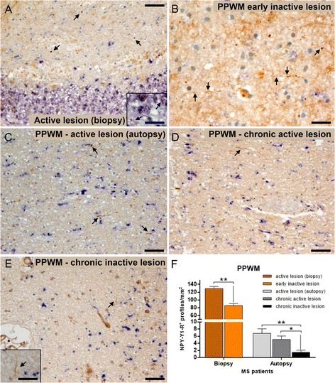
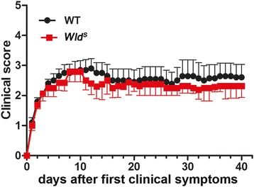
The highest numbers of axons undergoing Wallerian degeneration were found in the perilesional white matter of multiple sclerosis patients early in the disease course and with actively demyelinating lesions. Furthermore, Wallerian degeneration was more abundant in patients harboring chronic active as compared to chronic inactive lesions. No co-localization of neuropeptide Y-Y1 receptor, a bona fide immunohistochemical marker of Wallerian degeneration, with amyloid precursor protein, frequently used as an indicator of acute axonal transport disturbance, was observed in human and mouse tissue, indicating distinct axon-degenerative processes. Experimentally, a delay of Wallerian degeneration, as observed in Wld mice, did not result in a reduction of clinical disability or acute axonal damage in experimental autoimmune encephalomyelitis, further supporting that acute axonal damage as reflected by axonal transport disturbances does not share common molecular mechanisms with Wallerian degeneration. Furthermore, delaying Wallerian degeneration did not result in a net rescue of axons in late lesion stages of experimental autoimmune encephalomyelitis.
Our data indicate that in multiple sclerosis, ongoing demyelination in focal lesions is associated with axonal degeneration in the perilesional white matter, supporting a role for focal pathology in diffuse white matter damage. Also, our results suggest that interfering with Wallerian degeneration in inflammatory demyelination does not suffice to prevent acute axonal damage and finally axonal loss.
在进展性多发性硬化症中,轴突损伤和丢失在临床残疾的渐进性累积中起了很大作用。在此,我们评估了多发性硬化症患者脑组织中沃勒变性的程度与脱髓鞘病变活动的关系,并询问在炎症性脱髓鞘小鼠模型中,短暂阻断沃勒变性是否能减少轴突丢失和临床残疾。
采用免疫组织化学方法,在患有早期活动性脱髓鞘病变、慢性活动性病变和非活动性病变的多发性硬化症患者的斑块周围白质中,测定沃勒变性和急性轴突损伤。此外,我们使用沃勒变性缓慢(Wld )突变小鼠,研究了沃勒变性阻断对实验性自身免疫性脑脊髓炎的临床严重程度、炎症病理、急性轴突损伤和长期轴突丢失的影响。
在疾病病程早期且有活动性脱髓鞘病变的多发性硬化症患者的病变周围白质中,发现经历沃勒变性的轴突数量最多。此外,与慢性非活动性病变相比,患有慢性活动性病变的患者中沃勒变性更为明显。在人和小鼠组织中,未观察到沃勒变性的真正免疫组织化学标志物神经肽Y-Y1受体与经常用作急性轴突运输障碍指标的淀粉样前体蛋白共定位,这表明存在不同的轴突退行性过程。在实验中,如在Wld 小鼠中观察到的沃勒变性延迟,并未导致实验性自身免疫性脑脊髓炎的临床残疾或急性轴突损伤减少,进一步支持轴突运输障碍所反映的急性轴突损伤与沃勒变性没有共同的分子机制。此外,延迟沃勒变性并未导致实验性自身免疫性脑脊髓炎晚期病变阶段轴突的净挽救。
我们的数据表明,在多发性硬化症中,局灶性病变中持续的脱髓鞘与病变周围白质中的轴突变性相关,支持局灶性病理在弥漫性白质损伤中的作用。此外,我们的结果表明,在炎症性脱髓鞘中干扰沃勒变性不足以预防急性轴突损伤以及最终的轴突丢失。