Meguins Lucas C, Hidalgo Raquel C T, Spotti Antônio R, de Morais Dionei F
Department of Neurological Sciences, Division of Neurosurgery, Hospital de Base, Faculdade de Medicina de São José do Rio Preto (FAMERP/SP), São Paulo, Brazil.
Surg Neurol Int. 2017 Feb 20;8:25. doi: 10.4103/2152-7806.200577. eCollection 2017.
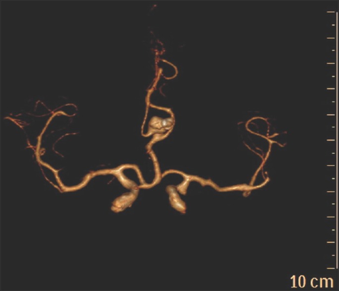
The azygos anterior cerebral artery (ACA) is an uncommon intracranial vascular anomaly of the circle of Willis. Identification of aneurysm from azygos ACA with anterior falcine meningioma is an extremely rare association. The aim of the present study is to report the case of an adult female with a ruptured aneurysm from azygos ACA in association with an anterior falcine meningioma.
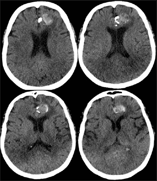
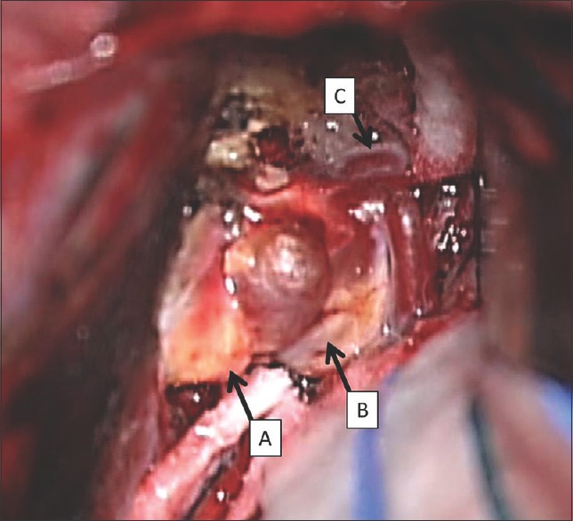
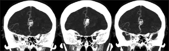
A 65-year-old female was admitted in the Emergency Department reporting sudden onset of severe headache. Computed tomography (CT) revealed an intracerebral hematoma and an expansive calcified lesion. AngioCT showed the presence of a large aneurysm in the distal portion of the azygos ACA. During the surgical procedure, it was possible to visualize the aneurysm in contact with an expansive lesion arising from the anterior third of the falx. Microsurgical clipping of the aneurysm was performed uneventfully and partial resection of the tumor was done. Histopathological analysis showed a fibrous meningioma. The patient was discharged home on the seventh postoperative day in good clinical conditions.
The association of aneurysm from azygos ACA and falcine meningioma is an extremely rare event and must be remembered when expansive masses are present in the vicinity of vascular lesions.
奇静脉大脑前动脉(ACA)是 Willis 环一种罕见的颅内血管异常。在奇静脉 ACA 合并大脑镰前部脑膜瘤时发现动脉瘤极为罕见。本研究旨在报告一例成年女性奇静脉 ACA 动脉瘤破裂并合并大脑镰前部脑膜瘤的病例。
一名 65 岁女性因突发剧烈头痛入住急诊科。计算机断层扫描(CT)显示脑内血肿和一个增大的钙化病变。CT 血管造影显示奇静脉 ACA 远端存在一个大动脉瘤。手术过程中,可以看到动脉瘤与起源于大脑镰前三分之一处的增大病变相接触。动脉瘤的显微夹闭手术顺利完成,肿瘤部分切除。组织病理学分析显示为纤维性脑膜瘤。患者术后第七天临床状况良好出院。
奇静脉 ACA 动脉瘤与大脑镰脑膜瘤的关联极为罕见,当血管病变附近出现增大肿块时必须予以考虑。