Voutilainen Merja H, De Lorenzo Francesca, Stepanova Polina, Bäck Susanne, Yu Li-Ying, Lindholm Päivi, Pörsti Eeva, Saarma Mart, Männistö Pekka T, Tuominen Raimo K
Division of Pharmacology and Pharmacotherapy, Faculty of Pharmacy, Viikki Biocenter, University of Helsinki, FIN-00014 Helsinki, Finland; Institute of Biotechnology, Viikki Biocenter, University of Helsinki, FIN-00014 Helsinki, Finland.
Institute of Biotechnology, Viikki Biocenter, University of Helsinki , FIN-00014 Helsinki, Finland.
eNeuro. 2017 Mar 13;4(1). doi: 10.1523/ENEURO.0117-16.2017. eCollection 2017 Jan-Feb.
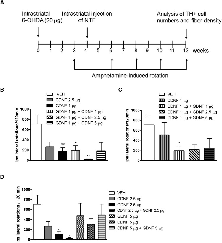
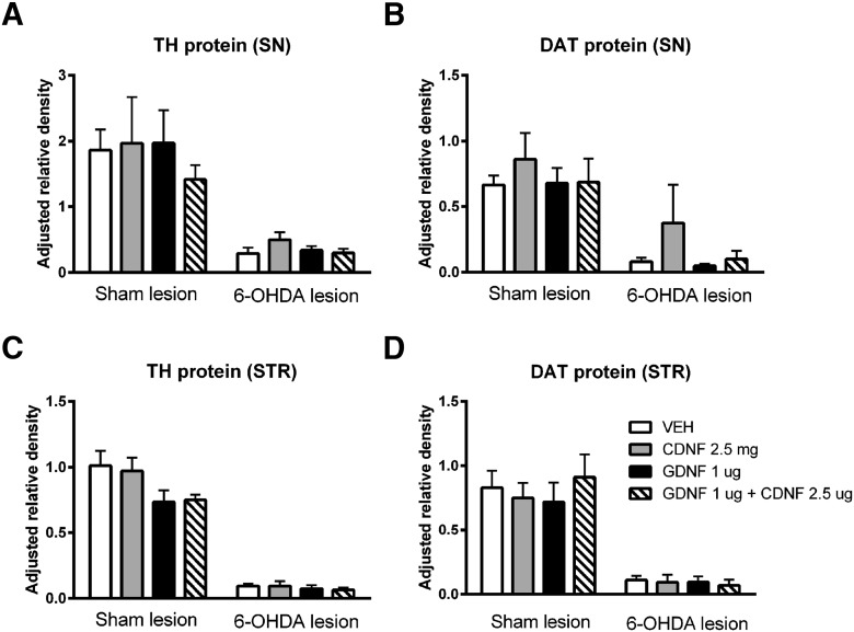
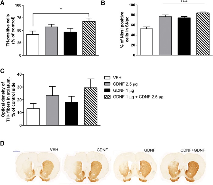
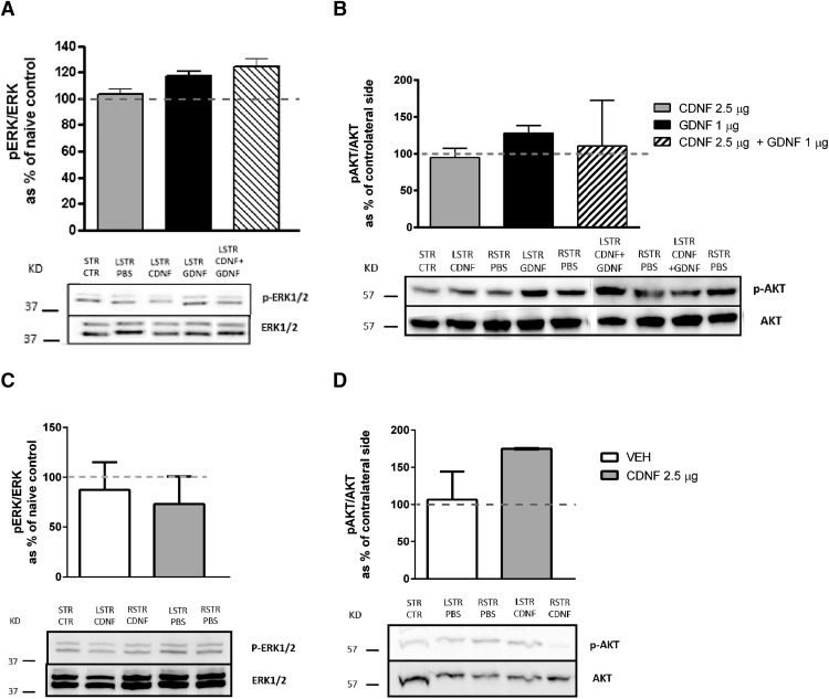
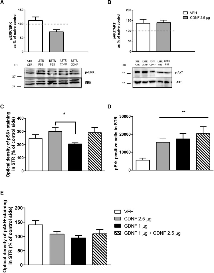
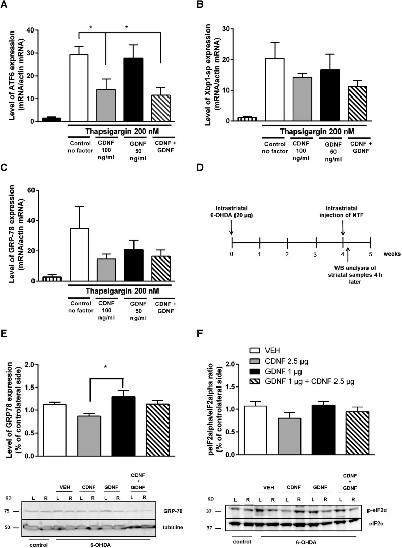
Parkinson's disease (PD) is a neurodegenerative disorder associated with a progressive loss of dopaminergic (DAergic) neurons of the substantia nigra (SN) and the accumulation of intracellular inclusions containing α-synuclein. Current therapies do not stop the progression of the disease, and the efficacy of these treatments wanes over time. Neurotrophic factors (NTFs) are naturally occurring proteins promoting the survival and differentiation of neurons and the maintenance of neuronal contacts. CDNF (cerebral dopamine NTF) and GDNF (glial cell line-derived NTF) are able to protect DAergic neurons against toxin-induced degeneration in experimental models of PD. Here, we report an additive neurorestorative effect of coadministration of CDNF and GDNF in the unilateral 6-hydroxydopamine (6-OHDA) lesion model of PD in rats. NTFs were given into the striatum four weeks after unilateral intrastriatal injection of 6-OHDA (20 µg). Amphetamine-induced (2.5 mg/kg, i.p.) rotational behavior was measured every two weeks. Number of tyrosine hydroxylase (TH)-positive cells from SN pars compacta (SNpc) and density of TH-positive fibers in the striatum were analyzed at 12 weeks after lesion. CDNF and GDNF alone restored the DAergic function, and one specific dose combination had an additive effect: CDNF (2.5µg) and GDNF (1µg) coadministration led to a stronger trophic effect relative to either of the single treatments alone. The additive effect may indicate different mechanism of action for the NTFs. Indeed, both NTFs activated the survival promoting PI3 kinase (PI3K)-Akt signaling pathway, but only CDNF decreased the expression level of tested endoplasmatic reticulum (ER) stress markers ATF6, glucose-regulated protein 78 (GRP78), and phosphorylation of eukaryotic initiation factor 2α subunit (eIF2α).
帕金森病(PD)是一种神经退行性疾病,与黑质(SN)中多巴胺能(DAergic)神经元的渐进性丧失以及含有α-突触核蛋白的细胞内包涵体的积累有关。目前的治疗方法无法阻止疾病的进展,并且这些治疗的效果会随着时间的推移而减弱。神经营养因子(NTFs)是天然存在的蛋白质,可促进神经元的存活和分化以及维持神经元联系。在帕金森病的实验模型中,脑源性神经营养因子(CDNF)和胶质细胞源性神经营养因子(GDNF)能够保护多巴胺能神经元免受毒素诱导的退化。在此,我们报告了在大鼠单侧6-羟基多巴胺(6-OHDA)损伤的帕金森病模型中,联合给予CDNF和GDNF的附加神经修复作用。在单侧纹状体内注射6-OHDA(20μg)四周后,将神经营养因子注入纹状体。每两周测量一次苯丙胺诱导的(2.5mg/kg,腹腔注射)旋转行为。在损伤后12周分析黑质致密部(SNpc)中酪氨酸羟化酶(TH)阳性细胞的数量以及纹状体中TH阳性纤维的密度。单独使用CDNF和GDNF可恢复多巴胺能功能,一种特定的剂量组合具有附加作用:联合给予CDNF(2.5μg)和GDNF(1μg)相对于单独的任何一种单一治疗均产生更强的营养作用。这种附加作用可能表明神经营养因子的作用机制不同。实际上,两种神经营养因子均激活了促进存活的磷脂酰肌醇-3激酶(PI3K)-Akt信号通路,但只有CDNF降低了所检测的内质网(ER)应激标志物活化转录因子6(ATF6)、葡萄糖调节蛋白78(GRP78)以及真核起始因子2α亚基(eIF2α)的磷酸化表达水平。