• 文献检索
  • 文档翻译
  • 深度研究
  • 学术资讯
  • Suppr Zotero 插件Zotero 插件
  • 邀请有礼
  • 套餐&价格
  • 历史记录
应用&插件
Suppr Zotero 插件Zotero 插件浏览器插件Mac 客户端Windows 客户端微信小程序
定价
高级版会员购买积分包购买API积分包
服务
文献检索文档翻译深度研究API 文档MCP 服务
关于我们
关于 Suppr公司介绍联系我们用户协议隐私条款
关注我们

Suppr 超能文献

核心技术专利:CN118964589B侵权必究
粤ICP备2023148730 号-1Suppr @ 2026

文献检索

告别复杂PubMed语法,用中文像聊天一样搜索,搜遍4000万医学文献。AI智能推荐,让科研检索更轻松。

立即免费搜索

文件翻译

保留排版,准确专业,支持PDF/Word/PPT等文件格式,支持 12+语言互译。

免费翻译文档

深度研究

AI帮你快速写综述,25分钟生成高质量综述,智能提取关键信息,辅助科研写作。

立即免费体验

使用对比增强型微型计算机断层扫描对雀鹰前肢内部解剖结构进行三维可视化。

Three-dimensional visualisation of the internal anatomy of the sparrowhawk () forelimb using contrast-enhanced micro-computed tomography.

作者信息

Bribiesca-Contreras Fernanda, Sellers William I

机构信息

Faculty of Science and Engineering, University of Manchester , Manchester , UK.

出版信息

PeerJ. 2017 Mar 15;5:e3039. doi: 10.7717/peerj.3039. eCollection 2017.

DOI:10.7717/peerj.3039
PMID:28316884
原文链接:https://pmc.ncbi.nlm.nih.gov/articles/PMC5356476/
Abstract

BACKGROUND

Gross dissection is a widespread method for studying animal anatomy, despite being highly destructive and time-consuming. X-ray computed tomography (CT) has been shown to be a non-destructive alternative for studying anatomical structures. However, in the past it has been limited to only being able to visualise mineralised tissues. In recent years, morphologists have started to use traditional X-ray contrast agents to allow the visualisation of soft tissue elements in the CT context. The aim of this project is to assess the ability of contrast-enhanced micro-CT (μCT) to construct a three-dimensional (3D) model of the musculoskeletal system of the bird wing and to quantify muscle geometry and any systematic changes due to shrinkage. We expect that this reconstruction can be used as an anatomical guide to the sparrowhawk wing musculature and form the basis of further biomechanical analysis of flight.

METHODS

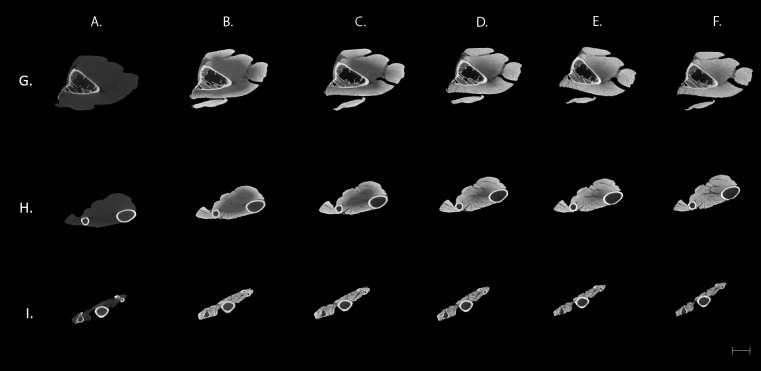
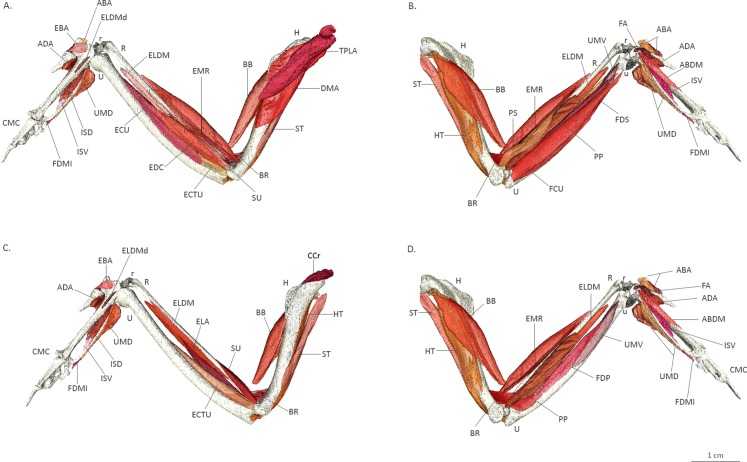
A 3% iodine-buffered formalin solution with a 25-day staining period was used to visualise the wing myology of the sparrowhawk (). μCT scans of the wing were taken over the staining period until full penetration of the forelimb musculature by iodine was reached. A 3D model was reconstructed by manually segmenting out the individual elements of the avian wing using 3D visualisation software.

RESULTS

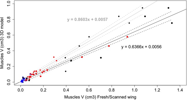
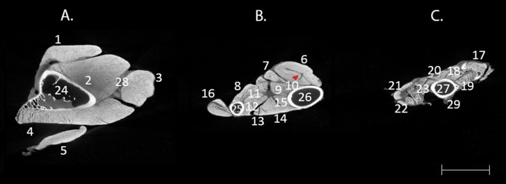
Different patterns of contrast were observed over the duration of the staining treatment with the best results occurring after 25 days of staining. Staining made it possible to visualise and identify different elements of the soft tissue of the wing. Finally, a 3D reconstruction of the musculoskeletal system of the sparrowhawk wing is presented and numerical data of muscle geometry is compared to values obtained by dissection.

DISCUSSION

Contrast-enhanced μCT allows the visualisation and identification of the wing myology of birds, including the smaller muscles in the hand, and provides a non-destructive way for quantifying muscle volume with an accuracy of 96.2%. By combining contrast-enhanced μCT with 3D visualisation techniques, it is possible to study the individual muscles of the forelimb in their original position and 3D design, which can be the basis of further biomechanical analysis. Because the stain can be washed out post analysis, this technique provides a means of obtaining quantitative muscle data from museum specimens non-destructively.

摘要

背景

大体解剖是研究动物解剖学的一种广泛应用的方法,尽管它具有高度的破坏性且耗时。X射线计算机断层扫描(CT)已被证明是研究解剖结构的一种非破坏性替代方法。然而,过去它仅限于能够可视化矿化组织。近年来,形态学家开始使用传统的X射线造影剂,以便在CT环境中可视化软组织成分。本项目的目的是评估对比增强型显微CT(μCT)构建鸟翼肌肉骨骼系统三维(3D)模型的能力,并量化肌肉几何形状以及由于收缩引起的任何系统性变化。我们期望这种重建能够用作雀鹰翅膀肌肉组织的解剖学指南,并构成进一步飞行生物力学分析的基础。

方法

使用3%碘缓冲福尔马林溶液,染色期为25天,以可视化雀鹰的翅膀肌肉学()。在染色期间对翅膀进行μCT扫描,直到碘完全穿透前肢肌肉组织。使用3D可视化软件手动分割出鸟类翅膀的各个元素,重建3D模型。

结果

在染色处理过程中观察到不同的对比模式,染色25天后效果最佳。染色使得可视化和识别翅膀软组织的不同元素成为可能。最后,展示了雀鹰翅膀肌肉骨骼系统的3D重建,并将肌肉几何形状的数值数据与通过解剖获得的值进行了比较。

讨论

对比增强型μCT能够可视化和识别鸟类的翅膀肌肉学,包括手部较小的肌肉,并提供一种非破坏性的方法来量化肌肉体积,准确率为96.2%。通过将对比增强型μCT与3D可视化技术相结合,可以在其原始位置和3D设计中研究前肢的各个肌肉,这可以作为进一步生物力学分析的基础。由于染色剂可以在分析后冲洗掉,该技术提供了一种从博物馆标本中无损获取定量肌肉数据的方法。

https://cdn.ncbi.nlm.nih.gov/pmc/blobs/209d/5356476/97eafc7da92f/peerj-05-3039-g004.jpg
https://cdn.ncbi.nlm.nih.gov/pmc/blobs/209d/5356476/d18806744e7a/peerj-05-3039-g001.jpg
https://cdn.ncbi.nlm.nih.gov/pmc/blobs/209d/5356476/985059d5f8f5/peerj-05-3039-g002.jpg
https://cdn.ncbi.nlm.nih.gov/pmc/blobs/209d/5356476/340b06df793a/peerj-05-3039-g003.jpg
https://cdn.ncbi.nlm.nih.gov/pmc/blobs/209d/5356476/97eafc7da92f/peerj-05-3039-g004.jpg

相似文献

1
Three-dimensional visualisation of the internal anatomy of the sparrowhawk () forelimb using contrast-enhanced micro-computed tomography.使用对比增强型微型计算机断层扫描对雀鹰前肢内部解剖结构进行三维可视化。
PeerJ. 2017 Mar 15;5:e3039. doi: 10.7717/peerj.3039. eCollection 2017.
2
Digital dissection of the pelvis and hindlimb of the red-legged running frog, , using Diffusible Iodine Contrast Enhanced computed microtomography (DICE CT).使用可扩散碘对比增强计算机显微断层扫描(DICE CT)对红腿奔蛙的骨盆和后肢进行数字解剖。
PeerJ. 2019 Jun 7;7:e7003. doi: 10.7717/peerj.7003. eCollection 2019.
3
A Quantitative and Comparative Analysis of the Muscle Architecture of the Forelimb Myology of Diurnal Birds of Prey (Order Accipitriformes and Falconiformes).昼行性猛禽(鹰形目和隼形目)前肢肌构筑的定量和比较分析。
Anat Rec (Hoboken). 2019 Oct;302(10):1808-1823. doi: 10.1002/ar.24195. Epub 2019 Jun 26.
4
Digital dissection - using contrast-enhanced computed tomography scanning to elucidate hard- and soft-tissue anatomy in the Common Buzzard Buteo buteo.数字解剖——利用增强对比计算机断层扫描来阐明普通鹰 Buteo buteo 的硬组织和软组织解剖结构。
J Anat. 2014 Apr;224(4):412-31. doi: 10.1111/joa.12153. Epub 2013 Dec 18.
5
Exploring polyoxometalates as non-destructive staining agents for contrast-enhanced microfocus computed tomography of biological tissues.探索多金属氧酸盐作为生物组织微焦点计算机断层扫描对比增强的非破坏性染色剂。
Acta Biomater. 2020 Mar 15;105:253-262. doi: 10.1016/j.actbio.2020.01.038. Epub 2020 Jan 26.
6
Enhancing CT imaging: A safe protocol to stain and de-stain rare fetal museum specimens using diffusible iodine-based staining (diceCT).增强 CT 成像:一种安全的方案,使用可扩散碘基染色剂(diceCT)对罕见胎儿博物馆标本进行染色和去染色。
J Anat. 2021 Jul;239(1):228-241. doi: 10.1111/joa.13410. Epub 2021 Mar 4.
7
Functional anatomy of the wing muscles of the Egyptian fruit bat (Rousettus aegyptiacus) using dissection and diceCT.利用解剖和数字容积成像技术对埃及果蝠(埃及果蝠)翼肌的功能解剖学研究
J Anat. 2025 May;246(5):685-695. doi: 10.1111/joa.14145. Epub 2024 Sep 30.
8
Iodine-Enhanced Micro-CT Imaging of Soft Tissue on the Example of Peripheral Nerve Regeneration.碘增强微 CT 成像在外周神经再生中的软组织应用。
Contrast Media Mol Imaging. 2019 Mar 27;2019:7483745. doi: 10.1155/2019/7483745. eCollection 2019.
9
Evaluation of contrasting techniques for X-ray imaging of velvet worms (Onychophora).评估 X 射线成像在有爪动物(Onychophora)中的对比技术。
J Microsc. 2018 Jun;270(3):343-358. doi: 10.1111/jmi.12688. Epub 2018 Feb 22.
10
Enhanced contrast in X-ray microtomographic images of the membranous labyrinth using different X-ray sources and scanning modes.使用不同的 X 射线源和扫描模式增强膜迷路的 X 射线微断层图像的对比度。
J Anat. 2018 Dec;233(6):770-782. doi: 10.1111/joa.12885. Epub 2018 Oct 2.

引用本文的文献

1
Morphlight theory inspired by raptors: musculoskeletal modeling and muscle control in Falco peregrinus wing flapping.受猛禽启发的形态光理论:游隼翅膀拍打中的肌肉骨骼建模与肌肉控制
Biol Open. 2025 Apr 15;14(4). doi: 10.1242/bio.061859. Epub 2025 Apr 10.
2
The SmARTR pipeline: A modular workflow for the cinematic rendering of 3D scientific imaging data.SmARTR工作流程:一种用于3D科学成像数据电影渲染的模块化工作流程。
iScience. 2024 Nov 23;27(12):111475. doi: 10.1016/j.isci.2024.111475. eCollection 2024 Dec 20.
3
Experimental assessment of diffusible iodine-based contrast-enhanced computed tomography (diceCT) protocols.

本文引用的文献

1
Functional anatomy of the pigeon hand (Columba livia): A muscle stimulation study.家鸽手部(岩鸽)的功能解剖:一项肌肉刺激研究。
J Morphol. 1995 Oct;226(1):33-45. doi: 10.1002/jmor.1052260104.
2
Flight feather attachment in rock pigeons (Columba livia): covert feathers and smooth muscle coordinate a morphing wing.原鸽(Columba livia)飞羽的附着:覆羽和平滑肌协调翅膀变形。
J Anat. 2016 Nov;229(5):631-656. doi: 10.1111/joa.12511. Epub 2016 Jun 20.
3
Diffusible iodine-based contrast-enhanced computed tomography (diceCT): an emerging tool for rapid, high-resolution, 3-D imaging of metazoan soft tissues.
基于弥散碘的对比增强计算机断层扫描(diceCT)方案的实验评估。
PeerJ. 2024 Sep 5;12:e17919. doi: 10.7717/peerj.17919. eCollection 2024.
4
Comparative muscle anatomy of the anuran pelvis and hindlimb in relation to locomotor mode.比较蛙类骨盆和后肢的肌肉解剖结构与运动方式的关系。
J Anat. 2024 Nov;245(5):751-774. doi: 10.1111/joa.14122. Epub 2024 Aug 9.
5
A histological and diceCT-derived 3D reconstruction of the avian visual thalamofugal pathway.鸟类视丘外侧通路的组织学和 DiceCT 三维重建。
Sci Rep. 2024 Apr 11;14(1):8447. doi: 10.1038/s41598-024-58788-z.
6
3D atlas of tinamou (Neornithes: Tinamidae) pectoral morphology: Implications for reconstructing the ancestral neornithine flight apparatus.平胸总目(新鸟亚纲:䳍科)胸肌形态的 3D 图谱:对重建祖先新鸟翅膀形态的启示。
J Anat. 2023 Nov;243(5):729-757. doi: 10.1111/joa.13919. Epub 2023 Jun 26.
7
Three-dimensional polygonal muscle modelling and line of action estimation in living and extinct taxa.活体和已灭绝分类群中的三维多角形肌肉建模和作用线估计。
Sci Rep. 2022 Mar 1;12(1):3358. doi: 10.1038/s41598-022-07074-x.
8
Hind foot drumming: Volumetric micro-computed tomography investigation of the hind limb musculature of three African mole-rat species (Bathyergidae).后脚击鼓:三种非洲鼹鼠科(Bathyergidae)后肢肌肉体积的微计算机断层扫描研究。
J Anat. 2022 Jan;240(1):23-33. doi: 10.1111/joa.13534. Epub 2021 Aug 9.
9
3D Muscle Architecture of the Pectoral Muscles of European Starling ().欧洲椋鸟胸肌的三维肌肉结构()。
Integr Org Biol. 2019 Feb 1;1(1):oby010. doi: 10.1093/iob/oby010. eCollection 2019.
10
ViceCT and whiceCT for simultaneous high-resolution visualization of craniofacial, brain and ventricular anatomy from micro-computed tomography.基于微计算机断层扫描的颅面、脑和脑室解剖的同时高分辨率可视化的 ViceCT 和 whiceCT
Sci Rep. 2020 Oct 30;10(1):18772. doi: 10.1038/s41598-020-75720-3.
基于扩散碘的对比增强计算机断层扫描(diceCT):一种用于后生动物软组织快速、高分辨率三维成像的新兴工具。
J Anat. 2016 Jun;228(6):889-909. doi: 10.1111/joa.12449. Epub 2016 Mar 11.
4
An investigation of the efficacy and mechanism of contrast-enhanced X-ray computed tomography utilizing iodine for large specimens through experimental and simulation approaches.通过实验和模拟方法对利用碘进行大样本对比增强X射线计算机断层扫描的有效性和机制进行研究。
BMC Physiol. 2015 Dec 21;15:5. doi: 10.1186/s12899-015-0019-3.
5
Frog tongue acts as muscle-powered adhesive tape.青蛙的舌头犹如肌肉驱动的胶带。
R Soc Open Sci. 2015 Sep 30;2(9):150333. doi: 10.1098/rsos.150333. eCollection 2015 Sep.
6
Three-dimensional non-destructive soft-tissue visualization with X-ray staining micro-tomography.利用X射线染色显微断层扫描技术进行三维无损软组织可视化。
Sci Rep. 2015 Sep 25;5:14088. doi: 10.1038/srep14088.
7
X-ray computed tomography of the anterior cruciate ligament and patellar tendon.前交叉韧带和髌腱的X线计算机断层扫描
Muscles Ligaments Tendons J. 2014 Jul 14;4(2):238-44. eCollection 2014 Apr.
8
Iodine vapor staining for atomic number contrast in backscattered electron and X-ray imaging.用于背散射电子和X射线成像中原子序数对比的碘蒸汽染色
Microsc Res Tech. 2014 Dec;77(12):1044-51. doi: 10.1002/jemt.22435. Epub 2014 Sep 15.
9
Digital dissection of the masticatory muscles of the naked mole-rat, Heterocephalus glaber (Mammalia, Rodentia).数字化解析裸鼹鼠(Mammalia,Rodentia)咀嚼肌。
PeerJ. 2014 Jun 17;2:e448. doi: 10.7717/peerj.448. eCollection 2014.
10
Volume shrinkage of bone, brain and muscle tissue in sample preparation for micro-CT and light sheet fluorescence microscopy (LSFM).样本制备过程中骨、脑和肌肉组织的体积收缩用于微计算机断层扫描和光片荧光显微镜(LSFM)。
Microsc Microanal. 2014 Aug;20(4):1208-17. doi: 10.1017/S1431927614001329. Epub 2014 Jun 25.