Di Pierdomenico Johnny, García-Ayuso Diego, Pinilla Isabel, Cuenca Nicolás, Vidal-Sanz Manuel, Agudo-Barriuso Marta, Villegas-Pérez María P
Departamento de Oftalmología, Facultad de Medicina, Universidad de Murcia and Instituto Murciano de Investigación Biosanitaria Virgen de la Arrixaca Murcia, Spain.
Instituto de Investigación Sanitaria Aragón, Aragon Health Sciences Institute, Lozano Blesa University Hospital Zaragoza, Spain.
Front Neuroanat. 2017 Mar 6;11:14. doi: 10.3389/fnana.2017.00014. eCollection 2017.
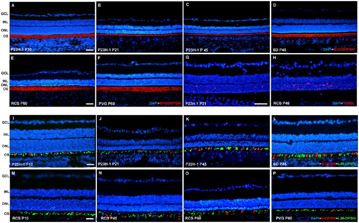
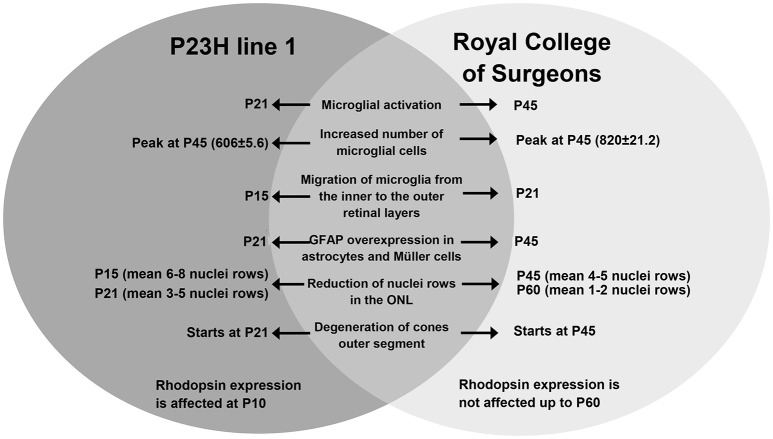
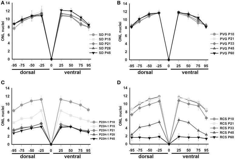
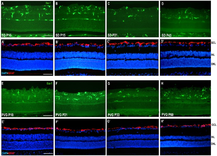
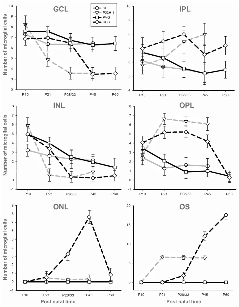
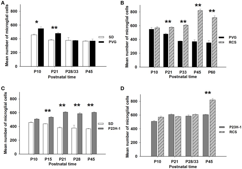
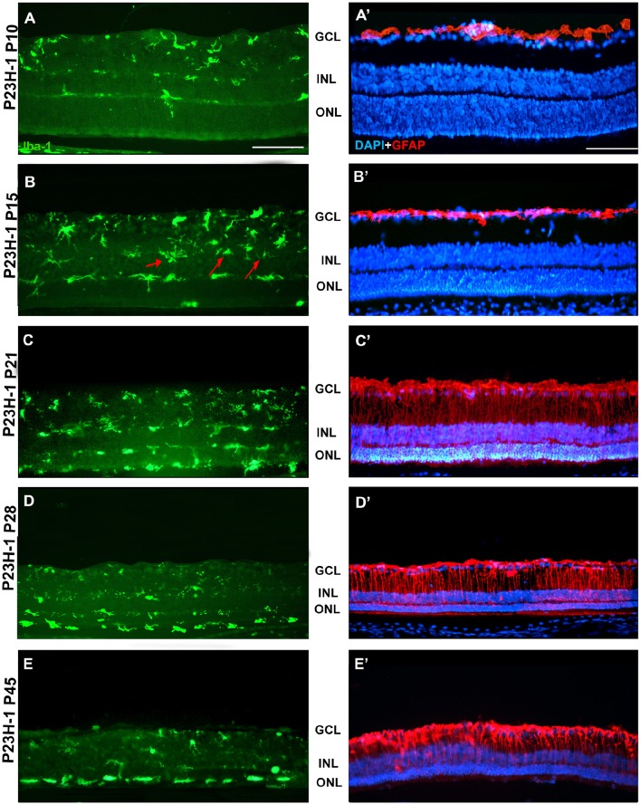
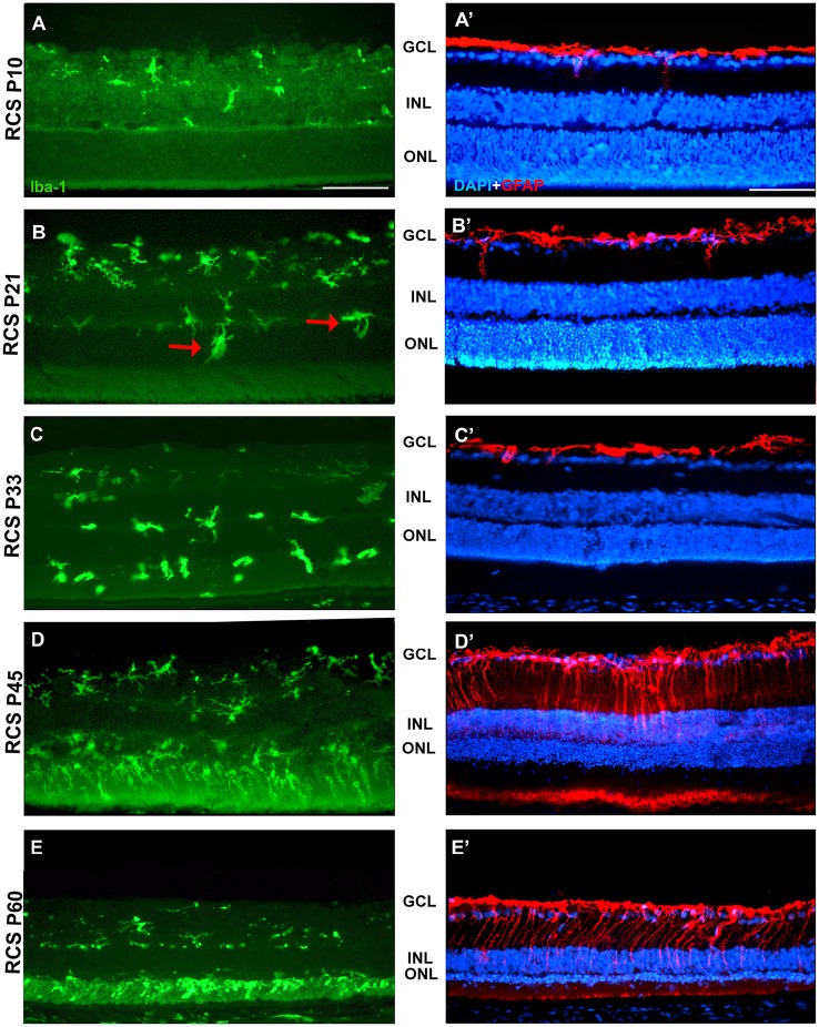
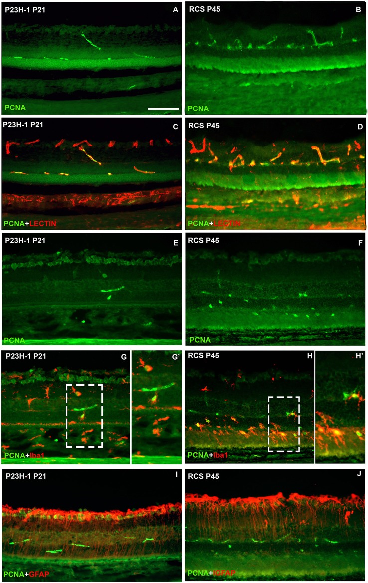
To study the course of photoreceptor cell death and macro and microglial reactivity in two rat models of retinal degeneration with different etiologies. Retinas from P23H-1 (rhodopsin mutation) and Royal College of Surgeon (RCS, pigment epithelium malfunction) rats and age-matched control animals (Sprague-Dawley and Pievald Viro Glaxo, respectively) were cross-sectioned at different postnatal ages (from P10 to P60) and rhodopsin, L/M- and S-opsin, ionized calcium-binding adapter molecule 1 (Iba1), glial fibrillary acid protein (GFAP), and proliferating cell nuclear antigen (PCNA) proteins were immunodetected. Photoreceptor nuclei rows and microglial cells in the different retinal layers were quantified. Photoreceptor degeneration starts earlier and progresses quicker in P23H-1 than in RCS rats. In both models, microglial cell activation occurs simultaneously with the initiation of photoreceptor death while GFAP over-expression starts later. As degeneration progresses, the numbers of microglial cells increase in the retina, but decreasing in the inner retina and increasing in the outer retina, more markedly in RCS rats. Interestingly, and in contrast with healthy animals, microglial cells reach the outer nuclei and outer segment layers. The higher number of microglial cells in dystrophic retinas cannot be fully accounted by intraretinal migration and PCNA immunodetection revealed microglial proliferation in both models but more importantly in RCS rats. The etiology of retinal degeneration determines the initiation and pattern of photoreceptor cell death and simultaneously there is microglial activation and migration, while the macroglial response is delayed. The actions of microglial cells in the degeneration cannot be explained only in the basis of photoreceptor death because they participate more actively in the RCS model. Thus, the retinal degeneration caused by pigment epithelium malfunction is more inflammatory and would probably respond better to interventions by inhibiting microglial cells.
研究两种不同病因的视网膜变性大鼠模型中光感受器细胞死亡过程以及巨噬细胞和小胶质细胞的反应性。对P23H-1(视紫红质突变)大鼠、皇家外科学院(RCS,色素上皮功能障碍)大鼠以及年龄匹配的对照动物(分别为Sprague-Dawley大鼠和Pievald Viro Glaxo大鼠)的视网膜在不同出生后年龄(从P10到P60)进行横切,并对视紫红质、L/M-和S-视蛋白、离子钙结合衔接分子1(Iba1)、胶质纤维酸性蛋白(GFAP)以及增殖细胞核抗原(PCNA)蛋白进行免疫检测。对不同视网膜层中的光感受器细胞核排数和小胶质细胞进行定量分析。P23H-1大鼠的光感受器变性比RCS大鼠更早开始且进展更快。在两种模型中,小胶质细胞激活与光感受器死亡的起始同时发生,而GFAP的过度表达则较晚开始。随着变性进展,视网膜中小胶质细胞数量增加,但在内层视网膜减少,在外层视网膜增加,在RCS大鼠中更为明显。有趣的是,与健康动物不同,小胶质细胞到达了外核层和外节层。营养不良视网膜中小胶质细胞数量的增加不能完全通过视网膜内迁移来解释,PCNA免疫检测显示两种模型中均有小胶质细胞增殖,但在RCS大鼠中更明显。视网膜变性的病因决定了光感受器细胞死亡的起始和模式,同时伴有小胶质细胞激活和迁移,而大胶质细胞反应则延迟。小胶质细胞在变性中的作用不能仅基于光感受器死亡来解释,因为它们在RCS模型中参与更为活跃。因此,由色素上皮功能障碍引起的视网膜变性炎症反应更强,可能对抑制小胶质细胞的干预反应更好。