Krysik Katarzyna, Dobrowolski Dariusz, Lyssek-Boron Anita, Jankowska-Szmul Judyta, Wylegala Edward A
Department of Ophthalmology with Pediatric Unit, St. Barbara Hospital, Trauma Center, Medykow Square 1, 41200 Sosnowiec, Poland.
Department of Ophthalmology with Pediatric Unit, St. Barbara Hospital, Trauma Center, Medykow Square 1, 41200 Sosnowiec, Poland; Clinical Departament of Ophthalmology, School of Medicine with the Division of Dentistry in Zabrze, Medical University of Silesia in Katowice, District Railway Hospital, Panewnicka 65 St., 40760 Katowice, Poland.
J Ophthalmol. 2017;2017:1582532. doi: 10.1155/2017/1582532. Epub 2017 Feb 23.
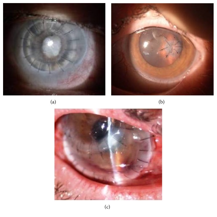
. To report the surgical approach, anatomical and functional results, and complications in the group of patients with corneal perforation. . 247 eyes with corneal perforation were operated on between January 2010 and July 2016. The three surgical procedures, dependent on size and location of perforation, were performed: full-sized penetrating keratoplasty, corneoscleral patch graft, and anterior lamellar keratoplasty. The eyes underwent the minimum 6-month follow-up visit. . Between January 2010 and July 2016, 247 surgeries were performed: 116 penetrating keratoplasties, 117 corneoscleral patch grafts, and 14 anterior lamellar keratoplasties. More than one procedure was necessary in 32 eyes. Final improvement of the visual acuity, within a gain of 2 or more lines with the Snellen test, was achieved in 56 operated eyes. To achieve better final visual acuity, 75 eyes required successive surgical treatment. Complications of the surgery comprised persistent epithelial defect, glaucoma or ocular hypertension, corneal oedema, graft melting, loose corneal sutures, reinfection, anterior synechiae and fibrinoid membranes, and endophthalmitis. In 26 eyes, the treatment failure was reported. . There is no one general-purpose surgical technique to treat corneal perforations. The complex nature of this pathology remains the individual, careful but also very distinct and multifactorial approach.
报告角膜穿孔患者组的手术方法、解剖和功能结果以及并发症。2010年1月至2016年7月期间,对247只角膜穿孔眼进行了手术。根据穿孔的大小和位置,实施了三种手术:全层穿透性角膜移植术、角膜巩膜补片移植术和前板层角膜移植术。这些眼睛接受了至少6个月的随访。2010年1月至2016年7月期间,共进行了247例手术:116例穿透性角膜移植术、117例角膜巩膜补片移植术和14例前板层角膜移植术。32只眼需要进行不止一种手术。56只手术眼的视力最终得到改善,用斯内伦视力表检查视力提高了2行或更多。为了获得更好的最终视力,75只眼需要进行连续的手术治疗。手术并发症包括持续性上皮缺损、青光眼或高眼压、角膜水肿、植片溶解、角膜缝线松动、再感染、虹膜前粘连和纤维蛋白样膜以及眼内炎。26只眼报告治疗失败。治疗角膜穿孔没有一种通用的手术技术。这种病理情况的复杂性仍然需要个体化、谨慎但也非常独特且多因素的方法。