Yoon Seo-Yeon, Kwon Soon-Gu, Kim Yong Ho, Yeo Ji-Hee, Ko Hyoung-Gon, Roh Dae-Hyun, Kaang Bong-Kiun, Beitz Alvin J, Lee Jang-Hern, Oh Seog Bae
1 Department of Brain and Cognitive Sciences, College of Natural Sciences, Pain Cognitive Function Research Center, Dental Research Institute, Seoul National University, Seoul, Republic of Korea.
2 Department of Neurobiology and Physiology School of Dentistry, Seoul National University, Seoul, Republic of Korea.
Mol Pain. 2017 Jan;13:1744806916688902. doi: 10.1177/1744806916688902.
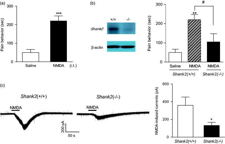
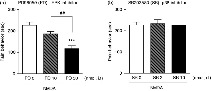
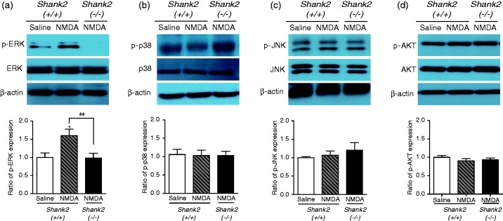
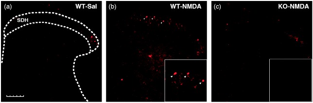
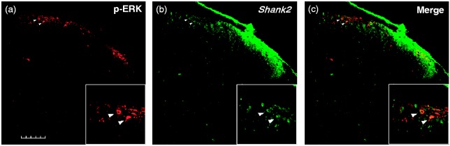
Background Self-injurious behaviors (SIBs) are devastating traits in autism spectrum disorder (ASD). Although deficits in pain sensation might be one of the contributing factors underlying the development of SIBs, the mechanisms have yet to be addressed. Recently, the Shank2 synaptic protein has been considered to be a key component in ASD, and mutations of SHANK2 gene induce the dysfunction of N-methyl-D-aspartate (NMDA) receptors, suggesting a link between Shank2 and NMDA receptors in ASD. Given that spinal NMDA receptors play a pivotal role in pain hypersensitivity, we investigated the possible role of Shank2 in nociceptive hypersensitivity by examining changes in spontaneous pain following intrathecal NMDA injection in S hank2-/- ( Shank2 knock-out, KO) mice. Results Intrathecal NMDA injection evoked spontaneous nociceptive behaviors. These NMDA-induced nociceptive responses were significantly reduced in Shank2 KO mice. We also observed a significant decrease of NMDA currents in the spinal dorsal horn of Shank2 KO mice. Subsequently, we examined whether mitogen-activated protein kinase or AKT signaling is involved in this reduced pain behavior in Shank2 KO mice because the NMDA receptor is closely related to these signaling molecules. Western blotting and immunohistochemistry revealed that spinally administered NMDA increased the expression of a phosphorylated form of extracellular signal-regulated kinase (p-ERK) which was significantly reduced in Shank2 KO mice. However, p38, JNK, or AKT were not changed by NMDA administration. The ERK inhibitor, PD98059, decreased NMDA-induced spontaneous pain behaviors in a dose-dependent manner in wild-type mice. Moreover, it was found that the NMDA-induced increase in p-ERK was primarily colocalized with Shank2 proteins in the spinal cord dorsal horn. Conclusion Shank2 protein is involved in spinal NMDA receptor-mediated pain, and mutations of Shank2 may suppress NMDA-ERK signaling in spinal pain transmission. This study provides new clues into the mechanisms underlying pain deficits associated with SIB and deserves further study in patients with ASD.
自伤行为(SIBs)是自闭症谱系障碍(ASD)中具有破坏性的特征。尽管痛觉缺陷可能是SIBs发生发展的促成因素之一,但其机制尚未得到阐明。最近,Shank2突触蛋白被认为是ASD的关键组成部分,并且SHANK2基因突变会导致N-甲基-D-天冬氨酸(NMDA)受体功能障碍,这表明ASD中Shank2与NMDA受体之间存在联系。鉴于脊髓NMDA受体在痛觉过敏中起关键作用,我们通过检查鞘内注射NMDA后Shank2基因敲除(Shank2-/-,KO)小鼠的自发疼痛变化,研究了Shank2在伤害性超敏反应中的可能作用。
鞘内注射NMDA诱发自发伤害性行为。这些NMDA诱导的伤害性反应在Shank2基因敲除小鼠中显著降低。我们还观察到Shank2基因敲除小鼠脊髓背角中NMDA电流显著减少。随后,我们研究了丝裂原活化蛋白激酶或AKT信号通路是否参与了Shank2基因敲除小鼠这种疼痛行为的减轻,因为NMDA受体与这些信号分子密切相关。蛋白质免疫印迹和免疫组织化学显示,鞘内注射NMDA可增加细胞外信号调节激酶磷酸化形式(p-ERK)的表达,而这种表达在Shank2基因敲除小鼠中显著降低。然而,p38、JNK或AKT在注射NMDA后没有变化。ERK抑制剂PD98059在野生型小鼠中以剂量依赖性方式降低了NMDA诱导的自发疼痛行为。此外,发现NMDA诱导的p-ERK增加主要与脊髓背角中的Shank2蛋白共定位。
Shank2蛋白参与脊髓NMDA受体介导的疼痛,并且Shank2突变可能抑制脊髓疼痛传递中的NMDA-ERK信号通路。本研究为与SIB相关的疼痛缺陷的潜在机制提供了新线索,值得在ASD患者中进一步研究。