Cheng Ni-Tao, Meng Hai, Ma Li-Feng, Zhang Liang, Yu Hao-Miao, Wang Zhen-Zhong, Guo Ai
Department of Orthopaedics, Beijing Friendship Hospital, Capital Medical University, Beijing 100050, P.R. China.
Int J Mol Med. 2017 May;39(5):1224-1232. doi: 10.3892/ijmm.2017.2934. Epub 2017 Mar 23.
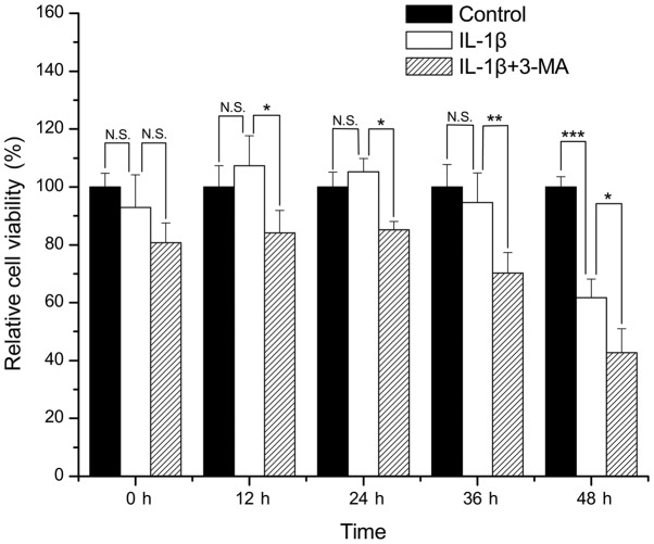
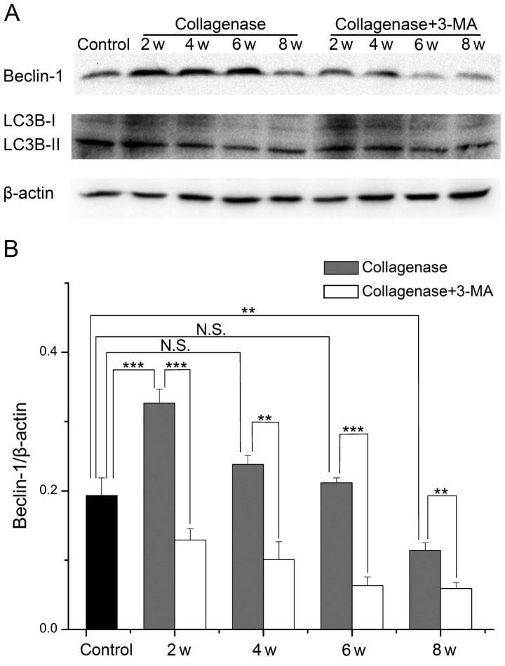
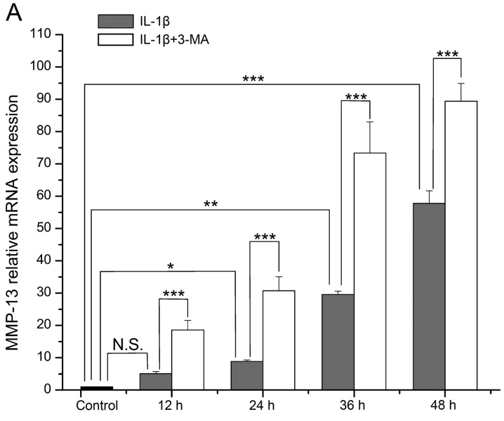
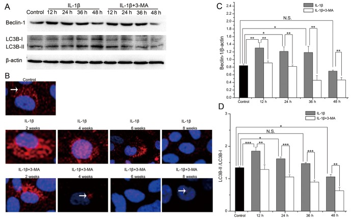
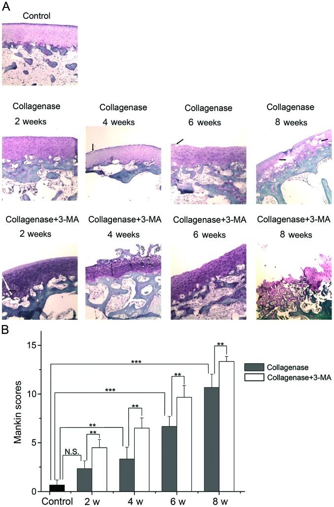
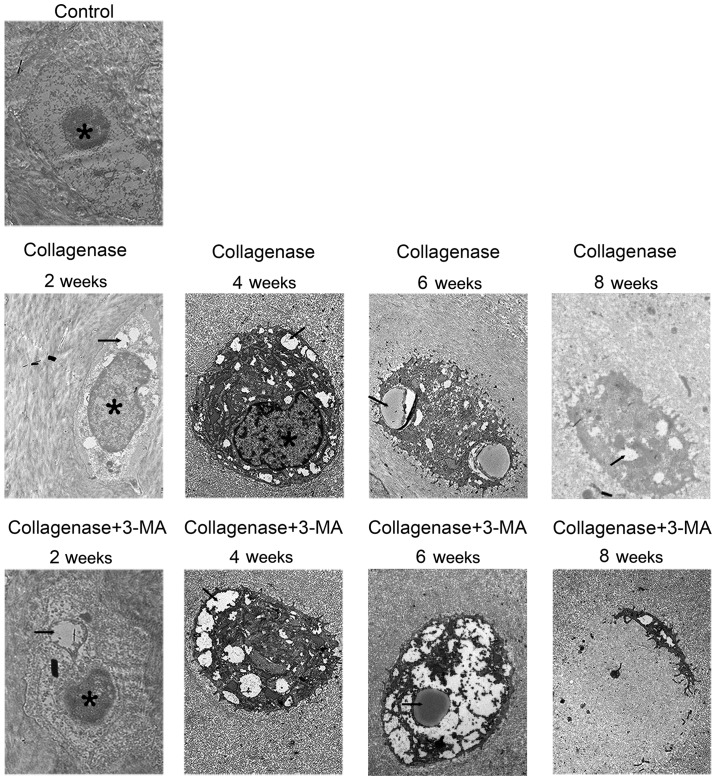
Accumulating evidence suggests that autophagy is closely related to the pathogenesis of osteoarthritis (OA). The aim of this study was to determine the changes in autophagy during the progression of OA and to elucidate the specific role of autophagy in OA. For this purpose, a cellular model of OA was generated by stimulating SW1353 cells with interleukin (IL)-1β and a rabbit model of OA was also established by an intra-articular injection of collagenase, followed by treatment with the autophagy specific inhibitor, 3-methyladenine (3-MA). Cell viability was analyzed by MTS assay, and the mRNA expression levels of matrix metalloproteinases (MMP)-13 and tissue inhibitor of metalloproteinase (TIMP)-1 were determined by RT-qPCR. Cartilage degeneration was examined under a light microscope, and autophagosome and chondrocyte degeneration was observed by transmission electron microscopy. The protein expression of Beclin-1 and light chain 3 (LC3)B was evaluated by western blot analysis and immunofluorescence staining. We found that the autophagy was enhanced during the early stages and was weakened during the late stages of experimental OA. The inhibition of autophagy by 3-MA significantly aggravated the degeneration of chondrocytes and cartilage in experimental OA. Our results thus determine the changes in autophagy during different stages of OA, as well as the role of impaired autophagy in the development of OA. Our data suggest that the regulation of autophagy may be a potential therapeutic strategy with which to attenuate OA.
越来越多的证据表明,自噬与骨关节炎(OA)的发病机制密切相关。本研究的目的是确定OA进展过程中自噬的变化,并阐明自噬在OA中的具体作用。为此,通过用白细胞介素(IL)-1β刺激SW1353细胞建立OA细胞模型,并用关节内注射胶原酶的方法建立OA兔模型,随后用自噬特异性抑制剂3-甲基腺嘌呤(3-MA)进行处理。通过MTS法分析细胞活力,通过RT-qPCR测定基质金属蛋白酶(MMP)-13和金属蛋白酶组织抑制剂(TIMP)-1的mRNA表达水平。在光学显微镜下检查软骨退变情况,通过透射电子显微镜观察自噬体和软骨细胞退变情况。通过蛋白质免疫印迹分析和免疫荧光染色评估Beclin-1和轻链3(LC3)B的蛋白表达。我们发现,在实验性OA的早期阶段自噬增强,而在晚期阶段自噬减弱。3-MA抑制自噬显著加重了实验性OA中软骨细胞和软骨的退变。因此,我们的结果确定了OA不同阶段自噬的变化,以及自噬受损在OA发展中的作用。我们的数据表明,自噬的调节可能是减轻OA的一种潜在治疗策略。