a School of Biological Sciences , Nanyang Technological University , Singapore.
b NTU Institute of Structural Biology , Nanyang Technological University , Singapore.
Nucleus. 2017 May 4;8(3):275-278. doi: 10.1080/19491034.2017.1287643. Epub 2017 Feb 7.
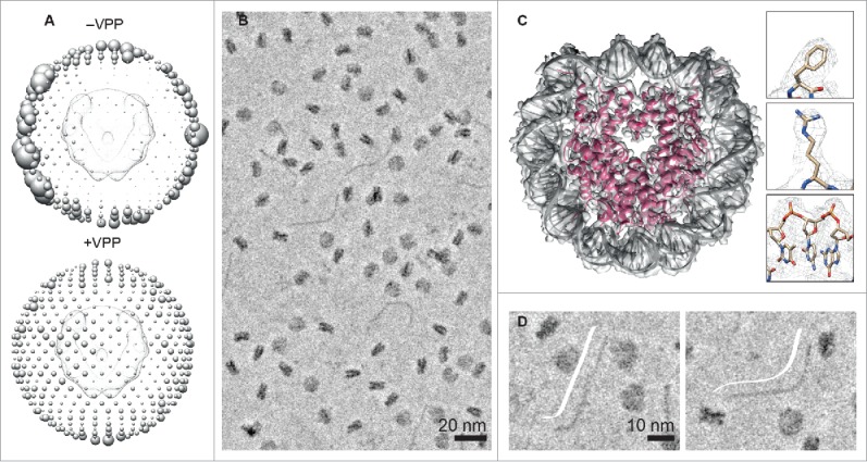
Contrast in electron cryo-microscopy (cryo-EM) is limited by the weak phase and radiation sensitive nature of biologic samples embedded in vitrified ice. We have recently shown that a new contrast enhancement technique utilizing the Volta phase plate can be combined with single particle analysis to determine the structure of a small chromatin complex, the nucleosome core particle, at near-atomic resolution. Here, we discuss advantages and limitations of the technique in terms of data collection, particle detection, and visualization of individual DNA molecules and higher-order chromatin structure.
电子冷冻透射显微镜(cryo-EM)的对比度受到限制,因为嵌入在玻璃态冰中的生物样本的相位很弱,对辐射也很敏感。我们最近表明,一种利用伏特相衬板的新技术可以与单颗粒分析结合使用,以确定一个小染色质复合物(核小体核心颗粒)的结构,达到近原子分辨率。在这里,我们将讨论该技术在数据收集、颗粒检测以及单个 DNA 分子和高级染色质结构可视化方面的优势和局限性。