Pathak Rekha, Aithal H P, Kinjavdekar P, Pawde A M, Tiwari A K, Sangeetha P, Tamilmahan P, Manzoor A B
Division of Veterinary Surgery, Indian Veterinary Research Institute, Izatnagar, Bareilly - 243 122, Uttar Pradesh, India.
Division of Standardization, Indian Veterinary Research Institute, Izatnagar, Bareilly - 243 122, Uttar Pradesh, India.
Vet World. 2017 Feb;10(2):163-169. doi: 10.14202/vetworld.2017.163-169. Epub 2017 Feb 8.
The aim of this study was to generate composite bone graft and investigate the rabbit fetal osteoblasts adhesion, proliferation and penetration on acellular matrices of cancellous bone.
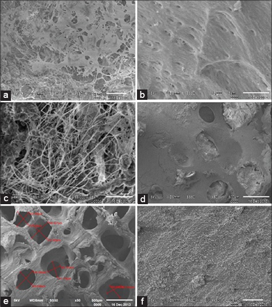
Acellular cancellous bone was prepared and developed as in the previous study with little modification. These matrices were decellularized by rapid freeze and thaw cycle. To remove the cell debris, they were then treated with hydrogen peroxide (3%) and ethanol to remove antigenic cellular and nuclear materials from the scaffold. Primary osteoblast cells were harvested from 20 to 22 days old rabbit fetal long and calvarial bone. These cells were cultured and characterized using a specific marker. The third passaged fetal osteoblast cells were then seeded on the scaffold and incubated for 14 days. The growth pattern of the cells was observed. Scanning electron microscope and hematoxylin and eosin staining were used to investigate cells proliferation.
The cells were found to be growing well on the surface of the scaffold and were also present in good numbers with the matrix filopodial extensions upto inside of the core of the tissue.
Thus, a viable composite scaffold of bone could be developed which has a great potential in the field of bone tissue engineering.
本研究旨在制备复合骨移植物,并研究兔胎儿成骨细胞在松质骨脱细胞基质上的黏附、增殖和穿透情况。
按照先前研究略作修改来制备和开发脱细胞松质骨。这些基质通过快速冻融循环进行脱细胞处理。为去除细胞碎片,随后用3%过氧化氢和乙醇处理,以从支架上去除抗原性细胞和核物质。从20至22日龄兔胎儿的长骨和颅骨中获取原代成骨细胞。使用特异性标志物对这些细胞进行培养和鉴定。然后将第三代传代的胎儿成骨细胞接种到支架上并孵育14天。观察细胞的生长模式。使用扫描电子显微镜和苏木精-伊红染色来研究细胞增殖情况。
发现细胞在支架表面生长良好,并且在基质中有大量丝状伪足延伸至组织核心内部。
因此,可以开发出一种可行的骨复合支架,其在骨组织工程领域具有巨大潜力。