Chen Fanfan, Li Zongyang, Weng Chengyin, Li Peng, Tu Lanbo, Chen Lei, Xie Wei, Li Ling
Neurosurgery Department, Guangzhou First People's Hospital, Guangzhou Medical University, Guangzhou, 510180, Guangdong, P. R. China.
Neurosurgery Department, Shenzhen Second People's Hospital, Shenzhen University, Shenzhen, 518000, Guangdong, P. R. China.
Chin J Cancer. 2017 Mar 27;36(1):34. doi: 10.1186/s40880-017-0201-z.
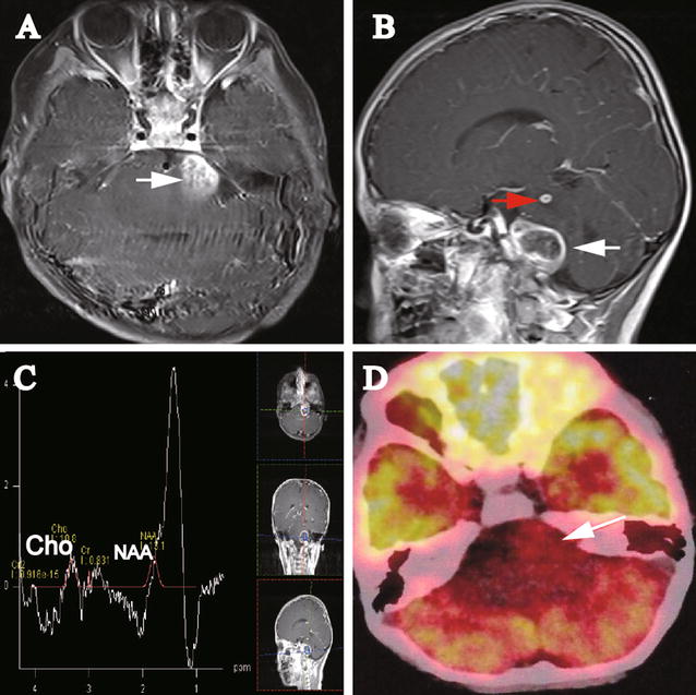
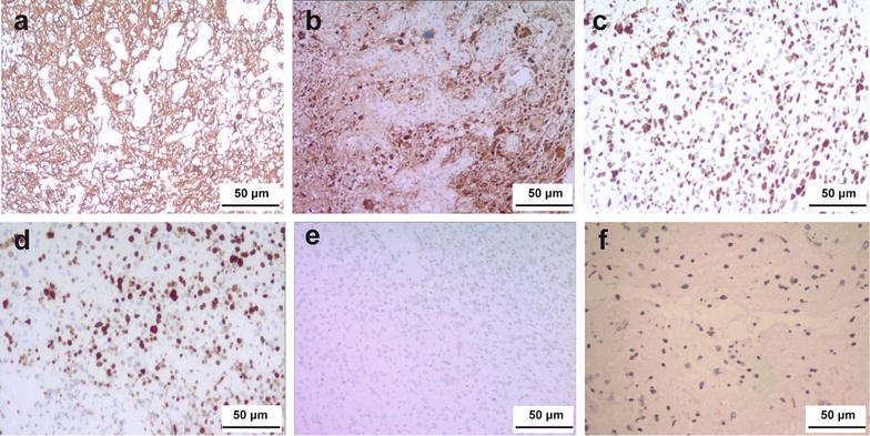
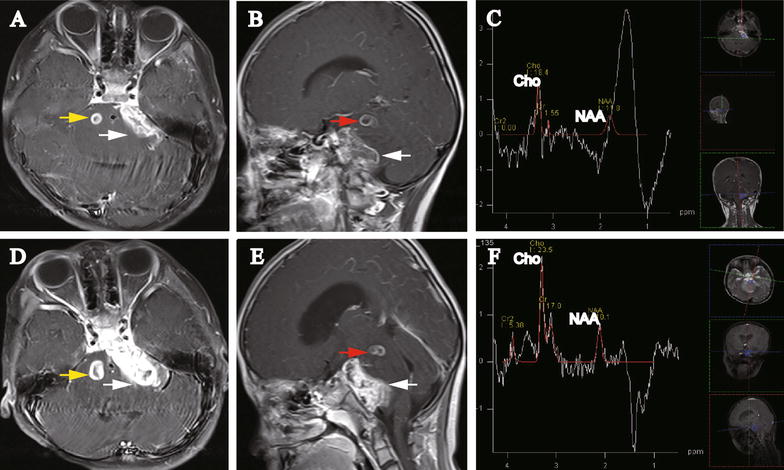
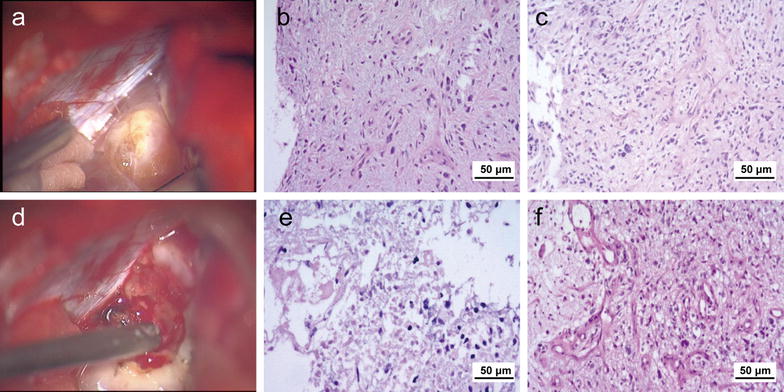
Multifocal pontine glioblastoma exhibiting an exophytic growth pattern in the cerebello-pontine angle (CPA) is rare. We present a case of a 5-year-old girl with consecutive neurological imaging and other clinical findings indicating progressive multifocal exophytic pontine glioblastoma. Three lesions were reported, of which two were initially presented, and one was developed 2 months later. One lesion demonstrated a progressing exophytic extension in the cistern of the left side of the CPA. The other two lesions were located and confined within the pons. Initial magnetic resonance imaging and positron emission tomography-computed tomography indicated low-grade glioma or inflammatory disease. However, 2 and 3 months later, subsequent magnetic resonance spectroscopy (MRS) displayed elevated choline and depressed N-acetyl aspartate peaks compared with the peaks on the initial MRS, indicating a high-grade glioma. Subtotal resection was performed for the CPA lesion. Histopathologic examination showed discrepant features of different parts of the CPA lesion. The patient received no further chemotherapy or radiotherapy and died 2 months after surgery. The multifocal and exophytic features of this case and the heterogeneous manifestations on neurological images were rare and confusing for both diagnosis and surgical decision-making. Our case report may contribute knowledge and helpful guidance for other medical doctors.
多灶性桥脑胶质母细胞瘤在小脑脑桥角(CPA)呈现外生性生长模式较为罕见。我们报告一例5岁女孩,其连续的神经影像学及其他临床检查结果提示为进行性多灶性外生性桥脑胶质母细胞瘤。报告有三个病灶,其中两个最初出现,一个在2个月后出现。一个病灶在左侧CPA脑池内呈现进行性外生性扩展。另外两个病灶位于桥脑内且局限于此。最初的磁共振成像和正电子发射断层扫描-计算机断层扫描显示为低级别胶质瘤或炎症性疾病。然而,在2个月和3个月后,后续的磁共振波谱(MRS)显示与初始MRS相比,胆碱峰升高,N-乙酰天门冬氨酸峰降低,提示为高级别胶质瘤。对CPA病灶进行了次全切除。组织病理学检查显示CPA病灶不同部位特征不一致。患者未接受进一步化疗或放疗,术后2个月死亡。该病例的多灶性和外生性特征以及神经影像学上的异质性表现罕见,对诊断和手术决策均造成困扰。我们的病例报告可能为其他医生提供知识和有益的指导。