Digby Genevieve C, Driver Helen S, Fitzpatrick Michael, Ropchan Glorianne, Parker Christopher M
Department of Medicine, Queen's University, Kingston, Ontario, Canada.
Department of Medicine, Queen's University, Kingston, Ontario, Canada; Sleep Disorders Laboratory, Kingston General Hospital, Kingston, Ontario, Canada.
Cardiol Res. 2011 Apr;2(2):51-57. doi: 10.4021/cr18w. Epub 2011 Mar 25.
Attempts to investigate the mechanisms by which continuous positive airway pressure (CPAP) therapy improves heart function in patients with obstructive sleep apnea (OSA) have been limited by the lack of non-invasive methods to assess cardiac performance. We used transthoracic electrical bioimpedance (TEB) to assess acute hemodynamic changes including heart rate (HR), stroke volume (SV), cardiac output (CO) and cardiac index (CI) during PAP titration in (1) post-operative cardiac surgery patients, (2) patients with severe OSA, and (3) normal healthy volunteers.
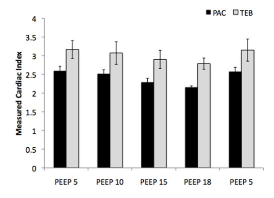
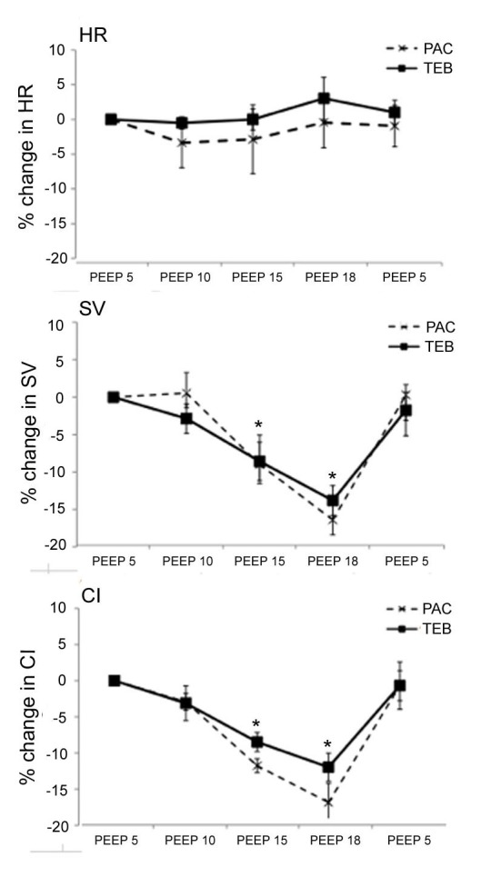
Post-operative cardiac surgery patients were studied via TEB and pulmonary artery catheter (PAC) during acute titration of positive end-expiratory pressure (PEEP) while mechanically ventilated. Patients with severe OSA were studied non-invasively by TEB during acute CPAP titration in supine stage 2 sleep, and normal subjects while awake and recumbent.
In post-operative cardiac surgery patients (n = 3), increasing PEEP to 18 cmHO significantly reduced SV and CI relative to baseline. There was no difference between TEB and PAC in terms of ability to assess variations in hemodynamic parameters. In patients with severe OSA (n = 3), CPAP titration to optimal pressure to alleviate obstructive apneas reduced HR, SV, CO and CI significantly compared to without CPAP. In three healthy subjects, maximal tolerated CPAP reduced SV and CO significantly compared to baseline.
Acute administration of CPAP causes a decrease in CO and CI, apparently a consequence of a reduction in SV. TEB appears to be an accurate and reproducible non-invasive method of detecting changes in hemodynamics.
由于缺乏评估心脏功能的非侵入性方法,探究持续气道正压通气(CPAP)治疗改善阻塞性睡眠呼吸暂停(OSA)患者心脏功能机制的尝试受到了限制。我们使用经胸电阻抗(TEB)来评估(1)心脏手术后患者、(2)重度OSA患者和(3)正常健康志愿者在PAP滴定期间的急性血流动力学变化,包括心率(HR)、每搏输出量(SV)、心输出量(CO)和心脏指数(CI)。
心脏手术后患者在机械通气时通过TEB和肺动脉导管(PAC)进行呼气末正压(PEEP)急性滴定研究。重度OSA患者在仰卧位2期睡眠期间进行急性CPAP滴定,通过TEB进行非侵入性研究,正常受试者在清醒和卧位时进行研究。
在心脏手术后患者(n = 3)中,将PEEP增加至18 cmH₂O相对于基线显著降低了SV和CI。在评估血流动力学参数变化的能力方面,TEB和PAC之间没有差异。在重度OSA患者(n = 3)中,与未使用CPAP相比,将CPAP滴定至最佳压力以缓解阻塞性呼吸暂停显著降低了HR、SV、CO和CI。在三名健康受试者中,与基线相比,最大耐受CPAP显著降低了SV和CO。
急性给予CPAP会导致CO和CI降低,这显然是SV降低的结果。TEB似乎是一种准确且可重复的检测血流动力学变化的非侵入性方法。