Zhou Ying, Yang Peng, Li Aili, Ye Xiaojun, Ren Shiyan, Li Xianlun
Department of Cardiology, China-Japan Friendship Hospital, Beijing 100029, P.R. China.
Biomed Rep. 2017 Feb;6(2):188-194. doi: 10.3892/br.2016.834. Epub 2016 Dec 29.
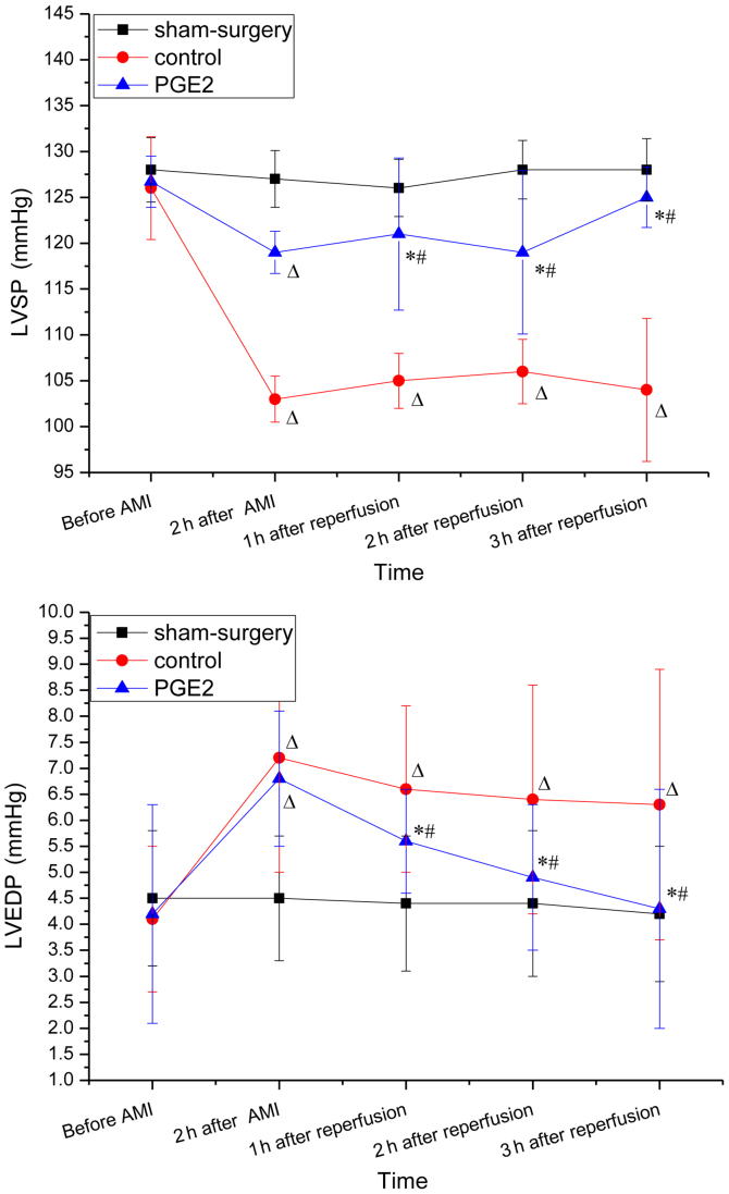
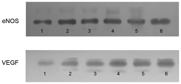
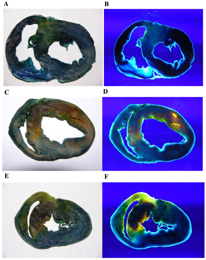
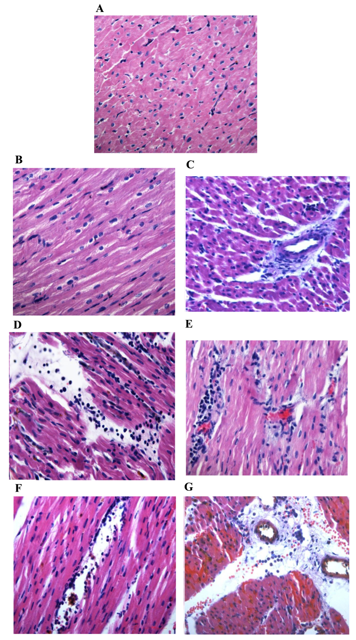
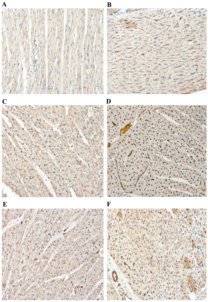
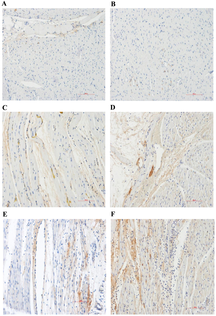
Prostaglandin E2 (PGE) has been demonstrated to attenuate cardiac ischemia-reperfusion (I/R) injury. However, the underlying mechanism of PGE in cardiac I/R injury remains unknown. Upregulated expression levels of vascular endothelial growth factor (VEGF) and endothelial nitric oxide synthase (eNOS) were reported in acute myocardial infarction (AMI), and were demonstrated to diminish I/R injury. In the current study the involvement of VEGF and eNOS in the myocardial protective effect of PGE were investigated in a catheter-based porcine model of AMI. Twenty-two Chinese miniature pigs were randomized into sham-surgery (n=6), control (n=8) and PGE (n=8) groups. PGE (1 µg/kg) was injected from 10 min prior to left anterior descending occlusion up to 1 h after reperfusion in the PGE group. Subsequently, the hemodynamic parameters were evaluated. Thioflavin-S and Evans Blue double staining were performed to evaluate the extent of the myocardial reperfusion area (RA) and no-reflow area (NRA). Immunohistochemical and western blot analysis were used to evaluate protein expression levels of VEGF and eNOS. Left ventricular (LV) systolic pressure significantly improved and LV end-diastolic pressure significantly decreased in the PGE group when compared with the control group 2 h after occlusion and 3 h after reperfusion (P<0.05, respectively). The RA and NRA were smaller in the PGE group than in the control group (P<0.05, respectively). Furthermore, PGE treatment increased the myocardial content of VEGF and eNOS when compared with the control group (P<0.05, respectively). Thus, the results of the present study demonstrate the cardio-protective mechanisms of PGE, which may protect the heart from I/R injury via enhancement of VEGF and eNOS expression levels.
前列腺素E2(PGE)已被证明可减轻心脏缺血再灌注(I/R)损伤。然而,PGE在心脏I/R损伤中的潜在机制仍不清楚。据报道,急性心肌梗死(AMI)中血管内皮生长因子(VEGF)和内皮型一氧化氮合酶(eNOS)的表达水平上调,并被证明可减轻I/R损伤。在本研究中,在基于导管的猪AMI模型中研究了VEGF和eNOS在PGE心肌保护作用中的作用。22只中国小型猪被随机分为假手术组(n=6)、对照组(n=8)和PGE组(n=8)。PGE组在左前降支闭塞前10分钟至再灌注后1小时注射PGE(1μg/kg)。随后,评估血流动力学参数。进行硫黄素-S和伊文思蓝双重染色以评估心肌再灌注区(RA)和无复流区(NRA)的范围。免疫组织化学和蛋白质印迹分析用于评估VEGF和eNOS的蛋白表达水平。与对照组相比,闭塞后2小时和再灌注后3小时,PGE组左心室(LV)收缩压显著改善,LV舒张末期压力显著降低(分别为P<0.05)。PGE组的RA和NRA均小于对照组(分别为P<0.05)。此外,与对照组相比,PGE治疗增加了心肌中VEGF和eNOS的含量(分别为P<0.05)。因此,本研究结果证明了PGE的心脏保护机制,其可能通过提高VEGF和eNOS表达水平来保护心脏免受I/R损伤。