Glogger Marius, Subota Ines, Pezzarossa Anna, Denecke Anna-Lena, Carrington Mark, Fenz Susanne F, Engstler Markus
Department of Cell and Developmental Biology, Biocenter, University of Würzburg, Würzburg, Germany.
Instituto de Medicina Molecular, Faculdade de Medicina, Universidade de Lisboa, Avenida Professor Egas Moniz, 1649-028 Lisboa, Portugal.
Exp Parasitol. 2017 Sep;180:13-18. doi: 10.1016/j.exppara.2017.03.010. Epub 2017 Mar 29.
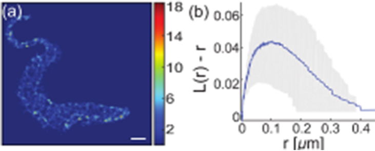
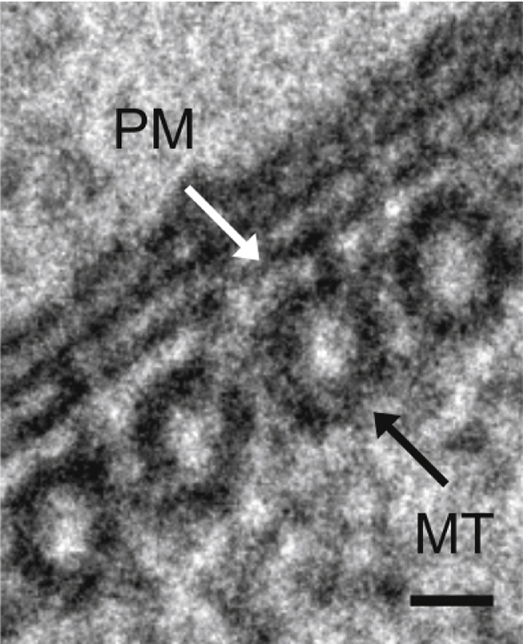
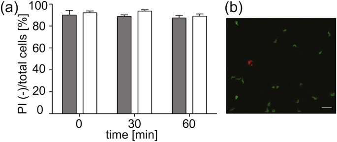
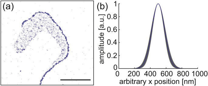
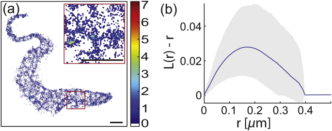
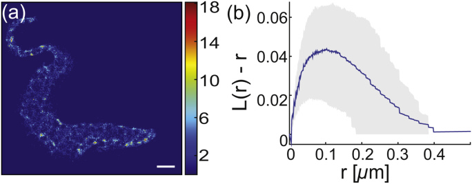
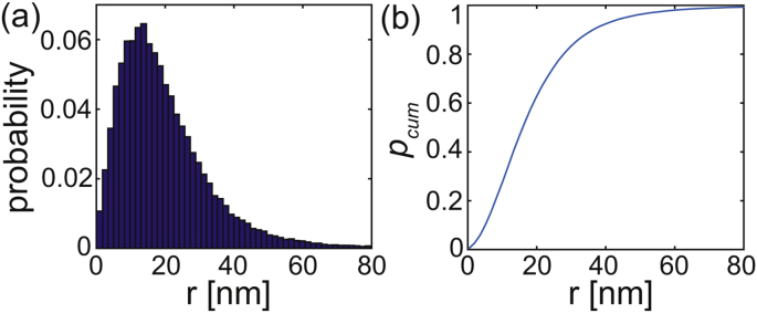
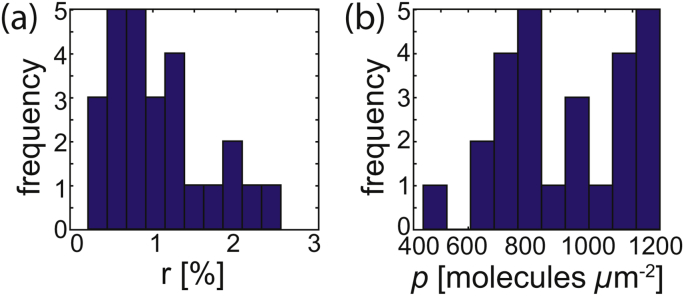
Research on trypanosomes as a model organism has provided a substantial contribution to a detailed understanding of basic cellular processes within the last few years. At the same time, major advances in super-resolution microscopy have been achieved, facilitating the resolution of biological structures in living cells at a scale of a few nm. However, the motility of trypanosomes has prevented access to high resolution microscopy of live cells. Here, we present a hydrogel based on poly(ethylene glycol) functionalized with either norbornene or thiol moieties for UV induced thiol-ene crosslinking for the embedding and imaging of live trypanosomes. The resulting gel exhibits low autofluorescence properties, immobilizes the cells efficiently on the nanometer scale and is compatible with cell viability for up to one hour at 24 °C. We applied super-resolution imaging to the inner plasma membrane leaflet using lipid-anchored eYFP as a probe. We find specific domains within the membrane where the fluorescence either accumulates or appears diluted rather than being homogenously distributed. Based on a Ripley's analysis, the size of the domains was determined to be r=170±5 nm and r>115±15 nm. We hypothesize that this structuring of the membrane is associated with the underlying cytoskeleton.
在过去几年里,将锥虫作为模式生物进行研究,为深入理解基本细胞过程做出了重大贡献。与此同时,超分辨率显微镜技术取得了重大进展,有助于在几纳米的尺度上解析活细胞中的生物结构。然而,锥虫的运动性使得难以对活细胞进行高分辨率显微镜观察。在此,我们展示了一种基于聚乙二醇的水凝胶,其用降冰片烯或硫醇部分进行功能化,用于紫外线诱导的硫醇-烯交联,以包埋和成像活锥虫。所得凝胶具有低自发荧光特性,能在纳米尺度上有效地固定细胞,并且在24℃下与细胞活力兼容长达一小时。我们使用脂质锚定的增强型黄色荧光蛋白(eYFP)作为探针,对内侧质膜小叶进行超分辨率成像。我们发现膜内存在特定区域,荧光在这些区域要么聚集要么显得稀释,而不是均匀分布。基于Ripley分析,确定这些区域的大小为r = 170±5纳米且r>1-15±15纳米。我们推测这种膜结构与潜在的细胞骨架有关。