Cui Lin, Rashdan Nabil A, Zhu Dongxing, Milne Elspeth M, Ajuh Paul, Milne Gillian, Helfrich Miep H, Lim Kelvin, Prasad Sai, Lerman Daniel A, Vesey Alex T, Dweck Marc R, Jenkins William S, Newby David E, Farquharson Colin, Macrae Vicky E
The Roslin Institute and Royal (Dick) School of Veterinary Studies, University of Edinburgh, Easter Bush, Edinburgh, United Kingdom.
Gemini Biosciences Ltd, Liverpool Science Park, Liverpool, United Kingdom.
J Cell Physiol. 2017 Nov;232(11):2985-2995. doi: 10.1002/jcp.25935. Epub 2017 May 24.
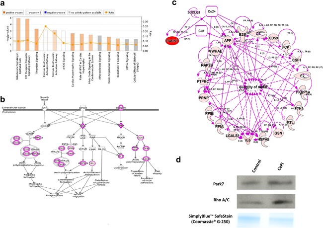
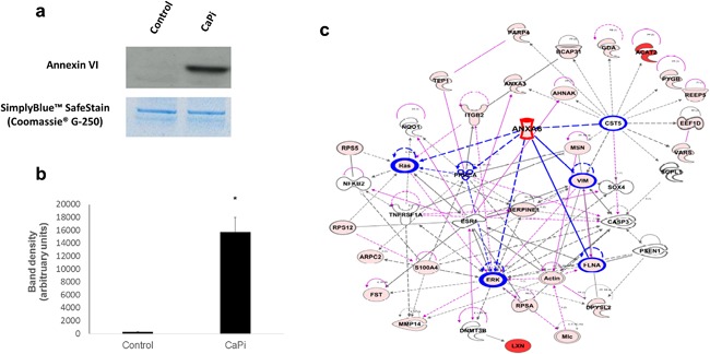
Patients with end-stage renal disease (ESRD) have elevated circulating calcium (Ca) and phosphate (Pi), and exhibit accelerated progression of calcific aortic valve disease (CAVD). We hypothesized that matrix vesicles (MVs) initiate the calcification process in CAVD. Ca induced rat valve interstitial cells (VICs) calcification at 4.5 mM (16.4-fold; p < 0.05) whereas Pi treatment alone had no effect. Ca (2.7 mM) and Pi (2.5 mM) synergistically induced calcium deposition (10.8-fold; p < 0.001) in VICs. Ca treatment increased the mRNA of the osteogenic markers Msx2, Runx2, and Alpl (p < 0.01). MVs were harvested by ultracentrifugation from VICs cultured with control or calcification media (containing 2.7 mM Ca and 2.5 mM Pi) for 16 hr. Proteomics analysis revealed the marked enrichment of exosomal proteins, including CD9, CD63, LAMP-1, and LAMP-2 and a concomitant up-regulation of the Annexin family of calcium-binding proteins. Of particular note Annexin VI was shown to be enriched in calcifying VIC-derived MVs (51.9-fold; p < 0.05). Through bioinformatic analysis using Ingenuity Pathway Analysis (IPA), the up-regulation of canonical signaling pathways relevant to cardiovascular function were identified in calcifying VIC-derived MVs, including aldosterone, Rho kinase, and metal binding. Further studies using human calcified valve tissue revealed the co-localization of Annexin VI with areas of MVs in the extracellular matrix by transmission electron microscopy (TEM). Together these findings highlight a critical role for VIC-derived MVs in CAVD. Furthermore, we identify calcium as a key driver of aortic valve calcification, which may directly underpin the increased susceptibility of ESRD patients to accelerated development of CAVD.
终末期肾病(ESRD)患者循环钙(Ca)和磷(Pi)水平升高,并表现出钙化性主动脉瓣疾病(CAVD)的加速进展。我们假设基质小泡(MVs)启动了CAVD中的钙化过程。4.5 mM的Ca诱导大鼠瓣膜间质细胞(VICs)钙化(16.4倍;p < 0.05),而单独的Pi处理没有效果。2.7 mM的Ca和2.5 mM的Pi协同诱导VICs中的钙沉积(10.8倍;p < 0.001)。Ca处理增加了成骨标志物Msx2、Runx2和Alpl的mRNA水平(p < 0.01)。通过超速离心从用对照或钙化培养基(含有2.7 mM Ca和2.5 mM Pi)培养16小时的VICs中收获MVs。蛋白质组学分析显示外泌体蛋白显著富集,包括CD9、CD63、LAMP-1和LAMP-2,以及钙结合蛋白膜联蛋白家族的伴随上调。特别值得注意的是,膜联蛋白VI在钙化的VIC衍生的MVs中富集(51.9倍;p < 0.05)。通过使用Ingenuity Pathway Analysis(IPA)的生物信息学分析,在钙化的VIC衍生的MVs中鉴定出与心血管功能相关的经典信号通路的上调,包括醛固酮、Rho激酶和金属结合。使用人钙化瓣膜组织的进一步研究通过透射电子显微镜(TEM)揭示了膜联蛋白VI与细胞外基质中MVs区域的共定位。这些发现共同突出了VIC衍生的MVs在CAVD中的关键作用。此外,我们确定钙是主动脉瓣钙化的关键驱动因素,这可能直接解释了ESRD患者对CAVD加速发展易感性增加的原因。