• 文献检索
  • 文档翻译
  • 深度研究
  • 学术资讯
  • Suppr Zotero 插件Zotero 插件
  • 邀请有礼
  • 套餐&价格
  • 历史记录
应用&插件
Suppr Zotero 插件Zotero 插件浏览器插件Mac 客户端Windows 客户端微信小程序
定价
高级版会员购买积分包购买API积分包
服务
文献检索文档翻译深度研究API 文档MCP 服务
关于我们
关于 Suppr公司介绍联系我们用户协议隐私条款
关注我们

Suppr 超能文献

核心技术专利:CN118964589B侵权必究
粤ICP备2023148730 号-1Suppr @ 2026

文献检索

告别复杂PubMed语法,用中文像聊天一样搜索,搜遍4000万医学文献。AI智能推荐,让科研检索更轻松。

立即免费搜索

文件翻译

保留排版,准确专业,支持PDF/Word/PPT等文件格式,支持 12+语言互译。

免费翻译文档

深度研究

AI帮你快速写综述,25分钟生成高质量综述,智能提取关键信息,辅助科研写作。

立即免费体验

利用新型瓣膜间质细胞系研究钙化性主动脉瓣疾病。

Exploiting novel valve interstitial cell lines to study calcific aortic valve disease.

机构信息

The Roslin Institute and Royal (Dick) School of Veterinary Studies, University of Edinburgh, Midlothian EH25 9RG, UK.

出版信息

Mol Med Rep. 2018 Feb;17(2):2100-2106. doi: 10.3892/mmr.2017.8163. Epub 2017 Nov 27.

DOI:10.3892/mmr.2017.8163
PMID:29207136
原文链接:https://pmc.ncbi.nlm.nih.gov/articles/PMC5783449/
Abstract

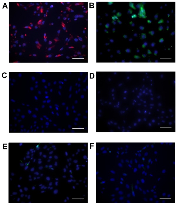
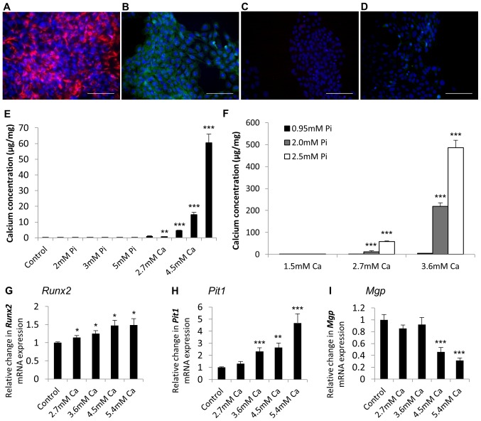
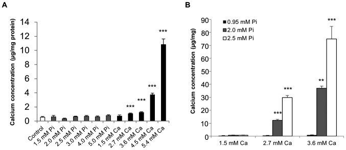
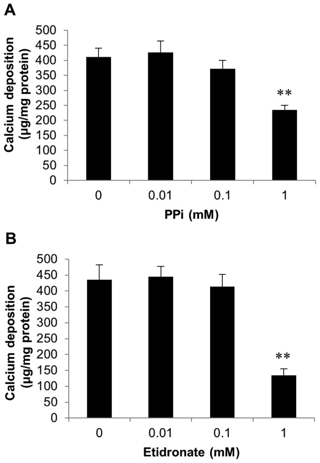
Calcific aortic valve disease (CAVD) involves progressive valve leaflet thickening and severe calcification, impairing leaflet motion. The in vitro calcification of primary rat, human, porcine and bovine aortic valve interstitial cells (VICs) is commonly employed to investigate CAVD mechanisms. However, to date, no published studies have utilised cell lines to investigate this process. The present study has therefore generated and evaluated the calcification potential of immortalized cell lines derived from sheep and rat VICs. Immortalised sheep (SAVIC) and rat (RAVIC) cell lines were produced by transduction with a recombinant lentivirus encoding the Simian virus (SV40) large and small T antigens (sheep), or large T antigen only (rat), which expressed markers of VICs (vimentin and α‑smooth muscle actin). Calcification was induced in the presence of calcium (Ca; 2.7 mM) in SAVICs (1.9 fold; P<0.001) and RAVICs (4.6 fold; P<0.01). Furthermore, a synergistic effect of calcium and phosphate was observed (2.7 mM Ca/2.0 mM Pi) on VIC calcification in the two cell lines (P<0.001). Analysis of SAVICs revealed significant increases in the mRNA expression of two key genes associated with vascular calcification in cells cultured under calcifying conditions, runt related transcription factor‑2 (RUNX2;1.3 fold; P<0.05 in 4.5 mM Ca) and sodium‑dependent phosphate transporter‑1 (PiT1; 1.2 fold; P<0.05 in 5.4 mM Ca). A concomitant decrease in the expression of the calcification inhibitor matrix Gla protein (MGP) was noted at 3.6 mM Ca (1.3 fold; P<0.01). Assessment of RAVICs revealed alterations in Runx2, Pit1 and Mgp mRNA expression levels (P<0.01). Furthermore, a significant reduction in calcification was observed in SAVICs following treatment with established calcification inhibitors, pyrophosphate (1.8 fold; P<0.01) and etidronate (3.2 fold; P<0.01). Overall, the present study demonstrated that the use of immortalised sheep and rat VIC cell lines is a convenient and cost effective system to investigate CAVD in vitro, and will make a useful contribution to increasing current understanding of the pathophysiological process.

摘要

钙化性主动脉瓣疾病 (CAVD) 涉及瓣叶进行性增厚和严重钙化,从而损害瓣叶运动。通常使用原代大鼠、人、猪和牛主动脉瓣膜间质细胞 (VICs) 的体外钙化来研究 CAVD 机制。然而,迄今为止,尚无研究利用细胞系来研究这一过程。因此,本研究生成并评估了源自绵羊和大鼠 VIC 的永生化细胞系的钙化潜力。通过转导含有编码猿猴病毒 (SV40) 大、小 T 抗原的重组慢病毒(绵羊),或仅大 T 抗原(大鼠),来产生永生化绵羊 (SAVIC) 和大鼠 (RAVIC) 细胞系,这些细胞系表达 VICs 的标志物(波形蛋白和α-平滑肌肌动蛋白)。在 SAVICs(增加 1.9 倍;P<0.001)和 RAVICs(增加 4.6 倍;P<0.01)中,在存在钙 (Ca; 2.7 mM) 的情况下诱导钙化。此外,在两种细胞系中观察到钙和磷酸盐的协同作用(2.7 mM Ca/2.0 mM Pi)对 VIC 钙化的影响(P<0.001)。对 SAVICs 的分析显示,在培养条件下,与血管钙化相关的两个关键基因的 mRNA 表达显著增加,即 runt 相关转录因子 2 (RUNX2; 在 4.5 mM Ca 时增加 1.3 倍;P<0.05) 和钠依赖性磷酸盐转运蛋白 1 (PiT1; 在 5.4 mM Ca 时增加 1.2 倍;P<0.05)。在 3.6 mM Ca 时,观察到钙化抑制剂基质 Gla 蛋白 (MGP) 的表达降低(降低 1.3 倍;P<0.01)。对 RAVICs 的评估显示,Runx2、Pit1 和 Mgp mRNA 表达水平发生改变(P<0.01)。此外,在 SAVICs 中,使用已建立的钙化抑制剂焦磷酸盐(降低 1.8 倍;P<0.01)和依替膦酸(降低 3.2 倍;P<0.01)处理后,钙化明显减少。总的来说,本研究表明,使用永生化绵羊和大鼠 VIC 细胞系是一种方便且具有成本效益的体外研究 CAVD 的系统,将为增加对病理生理过程的现有理解做出有用贡献。

https://cdn.ncbi.nlm.nih.gov/pmc/blobs/7ea5/5783449/9f05b64225f3/MMR-17-02-2100-g04.jpg
https://cdn.ncbi.nlm.nih.gov/pmc/blobs/7ea5/5783449/1452f1388246/MMR-17-02-2100-g00.jpg
https://cdn.ncbi.nlm.nih.gov/pmc/blobs/7ea5/5783449/76db5525b385/MMR-17-02-2100-g01.jpg
https://cdn.ncbi.nlm.nih.gov/pmc/blobs/7ea5/5783449/77c81cdf51bb/MMR-17-02-2100-g02.jpg
https://cdn.ncbi.nlm.nih.gov/pmc/blobs/7ea5/5783449/fe223194b1b9/MMR-17-02-2100-g03.jpg
https://cdn.ncbi.nlm.nih.gov/pmc/blobs/7ea5/5783449/9f05b64225f3/MMR-17-02-2100-g04.jpg

相似文献

1
Exploiting novel valve interstitial cell lines to study calcific aortic valve disease.利用新型瓣膜间质细胞系研究钙化性主动脉瓣疾病。
Mol Med Rep. 2018 Feb;17(2):2100-2106. doi: 10.3892/mmr.2017.8163. Epub 2017 Nov 27.
2
End stage renal disease-induced hypercalcemia may promote aortic valve calcification via Annexin VI enrichment of valve interstitial cell derived-matrix vesicles.终末期肾病引起的高钙血症可能通过膜联蛋白VI富集瓣膜间质细胞衍生的基质小泡促进主动脉瓣钙化。
J Cell Physiol. 2017 Nov;232(11):2985-2995. doi: 10.1002/jcp.25935. Epub 2017 May 24.
3
Transforming growth factor-β1 promotes fibrosis but attenuates calcification of valvular tissue applied as a three-dimensional calcific aortic valve disease model.转化生长因子-β1 促进纤维化,但可减轻作为三维钙化主动脉瓣疾病模型的瓣膜组织的钙化。
Am J Physiol Heart Circ Physiol. 2020 Nov 1;319(5):H1123-H1141. doi: 10.1152/ajpheart.00651.2019. Epub 2020 Sep 28.
4
Simulation of early calcific aortic valve disease in a 3D platform: A role for myofibroblast differentiation.3D平台中早期钙化性主动脉瓣疾病的模拟:肌成纤维细胞分化的作用。
J Mol Cell Cardiol. 2016 May;94:13-20. doi: 10.1016/j.yjmcc.2016.03.004. Epub 2016 Mar 17.
5
Isolation and Characterization of Primary Rat Valve Interstitial Cells: A New Model to Study Aortic Valve Calcification.原代大鼠瓣膜间质细胞的分离与鉴定:一种研究主动脉瓣钙化的新模型。
J Vis Exp. 2017 Nov 20(129):56126. doi: 10.3791/56126.
6
Comparison of calcification potential of valvular interstitial cells isolated from individual aortic valve cusps.从个体主动脉瓣叶分离的瓣膜间质细胞钙化潜能的比较。
Cardiovasc Pathol. 2016 May-Jun;25(3):185-194. doi: 10.1016/j.carpath.2015.12.002. Epub 2015 Dec 30.
7
MicroRNA-22 promoted osteogenic differentiation of valvular interstitial cells by inhibiting CAB39 expression during aortic valve calcification.MicroRNA-22 通过抑制主动脉瓣钙化过程中 CAB39 的表达促进瓣膜间质细胞的成骨分化。
Cell Mol Life Sci. 2022 Feb 21;79(3):146. doi: 10.1007/s00018-022-04177-6.
8
Dipeptidyl Peptidase-4 Induces Aortic Valve Calcification by Inhibiting Insulin-Like Growth Factor-1 Signaling in Valvular Interstitial Cells.二肽基肽酶-4 通过抑制瓣膜间质细胞中的胰岛素样生长因子-1 信号诱导主动脉瓣钙化。
Circulation. 2017 May 16;135(20):1935-1950. doi: 10.1161/CIRCULATIONAHA.116.024270. Epub 2017 Feb 8.
9
Valvular interstitial cells suppress calcification of valvular endothelial cells.瓣膜间质细胞抑制瓣膜内皮细胞的钙化。
Atherosclerosis. 2015 Sep;242(1):251-260. doi: 10.1016/j.atherosclerosis.2015.07.008. Epub 2015 Jul 17.
10
New calcification model for intact murine aortic valves.完整鼠主动脉瓣新钙化模型。
J Mol Cell Cardiol. 2021 Jul;156:95-104. doi: 10.1016/j.yjmcc.2021.03.003. Epub 2021 Mar 18.

引用本文的文献

1
Metformin ameliorates valve interstitial cell calcification by promoting autophagic flux.二甲双胍通过促进自噬流改善瓣膜间质细胞钙化。
Sci Rep. 2023 Dec 5;13(1):21435. doi: 10.1038/s41598-023-47774-6.
2
Challenges of aortic valve tissue culture - maintenance of viability and extracellular matrix in the pulsatile dynamic microphysiological system.主动脉瓣组织培养的挑战——在搏动动态微生理系统中维持活力和细胞外基质
J Biol Eng. 2023 Sep 28;17(1):60. doi: 10.1186/s13036-023-00377-1.
3
Multiscale structure and function of the aortic valve apparatus.

本文引用的文献

1
End stage renal disease-induced hypercalcemia may promote aortic valve calcification via Annexin VI enrichment of valve interstitial cell derived-matrix vesicles.终末期肾病引起的高钙血症可能通过膜联蛋白VI富集瓣膜间质细胞衍生的基质小泡促进主动脉瓣钙化。
J Cell Physiol. 2017 Nov;232(11):2985-2995. doi: 10.1002/jcp.25935. Epub 2017 May 24.
2
Bone Morphogenetic Protein Signaling Is Required for Aortic Valve Calcification.骨形态发生蛋白信号传导是主动脉瓣钙化所必需的。
Arterioscler Thromb Vasc Biol. 2016 Jul;36(7):1398-405. doi: 10.1161/ATVBAHA.116.307526. Epub 2016 May 19.
3
Calcific aortic stenosis.
主动脉瓣装置的多尺度结构与功能
Physiol Rev. 2024 Oct 1;104(4):1487-1532. doi: 10.1152/physrev.00038.2022. Epub 2023 Sep 21.
4
Uremic Toxin Lanthionine Induces Endothelial Cell Mineralization In Vitro.尿毒症毒素羊毛硫氨酸在体外诱导内皮细胞矿化。
Biomedicines. 2022 Feb 14;10(2):444. doi: 10.3390/biomedicines10020444.
5
The genetic consequences of dog breed formation-Accumulation of deleterious genetic variation and fixation of mutations associated with myxomatous mitral valve disease in cavalier King Charles spaniels.犬种形成的遗传后果-积累有害遗传变异和固定与可卡颇犬二尖瓣黏液样变性相关的突变。
PLoS Genet. 2021 Sep 2;17(9):e1009726. doi: 10.1371/journal.pgen.1009726. eCollection 2021 Sep.
6
Expression of Calcification and Extracellular Matrix Genes in the Cardiovascular System of the Healthy Domestic Sheep ().健康家养绵羊心血管系统中钙化和细胞外基质基因的表达()。
Front Genet. 2020 Sep 8;11:919. doi: 10.3389/fgene.2020.00919. eCollection 2020.
7
MicroRNA-214 promotes the calcification of human aortic valve interstitial cells through the acceleration of inflammatory reactions with activated MyD88/NF-κB signaling.微小 RNA-214 通过加速激活的 MyD88/NF-κB 信号转导的炎症反应促进人主动脉瓣间质细胞的钙化。
Clin Res Cardiol. 2019 Jun;108(6):691-702. doi: 10.1007/s00392-018-1398-9. Epub 2018 Dec 5.
8
Scanning electron microscopy for blood micro-crystals in aortic stenosis patients.主动脉瓣狭窄患者血液微晶体的扫描电子显微镜检查。
PLoS One. 2018 Aug 23;13(8):e0202282. doi: 10.1371/journal.pone.0202282. eCollection 2018.
9
Isolation and Characterization of Primary Rat Valve Interstitial Cells: A New Model to Study Aortic Valve Calcification.原代大鼠瓣膜间质细胞的分离与鉴定:一种研究主动脉瓣钙化的新模型。
J Vis Exp. 2017 Nov 20(129):56126. doi: 10.3791/56126.
钙化性主动脉瓣狭窄。
Nat Rev Dis Primers. 2016 Mar 3;2:16006. doi: 10.1038/nrdp.2016.6.
4
A novel role for the mineralocorticoid receptor in glucocorticoid driven vascular calcification.盐皮质激素受体在糖皮质激素驱动的血管钙化中的新作用。
Vascul Pharmacol. 2016 Nov;86:87-93. doi: 10.1016/j.vph.2016.04.005. Epub 2016 May 3.
5
Ablation of the androgen receptor from vascular smooth muscle cells demonstrates a role for testosterone in vascular calcification.从血管平滑肌细胞中去除雄激素受体证明了睾酮在血管钙化中的作用。
Sci Rep. 2016 Apr 20;6:24807. doi: 10.1038/srep24807.
6
Dual Effects of Bisphosphonates on Ectopic Skin and Vascular Soft Tissue Mineralization versus Bone Microarchitecture in a Mouse Model of Generalized Arterial Calcification of Infancy.双膦酸盐对婴儿全身性动脉钙化小鼠模型中异位皮肤和血管软组织矿化与骨微结构的双重影响
J Invest Dermatol. 2016 Jan;136(1):275-283. doi: 10.1038/JID.2015.377.
7
An In Vitro Murine Model of Vascular Smooth Muscle Cell Mineralization.血管平滑肌细胞矿化的体外小鼠模型
Methods Mol Biol. 2016;1397:209-220. doi: 10.1007/978-1-4939-3353-2_14.
8
Calcification in Aortic Stenosis: The Skeleton Key.主动脉瓣狭窄中的钙化:关键所在。
J Am Coll Cardiol. 2015 Aug 4;66(5):561-77. doi: 10.1016/j.jacc.2015.05.066.
9
Modulation of human valve interstitial cell phenotype and function using a fibroblast growth factor 2 formulation.使用成纤维细胞生长因子2制剂调节人瓣膜间质细胞表型和功能。
PLoS One. 2015 Jun 4;10(6):e0127844. doi: 10.1371/journal.pone.0127844. eCollection 2015.
10
Aortic stenosis begets aortic stenosis: between a rock and a hard place?主动脉狭窄引发主动脉狭窄:进退两难?
Heart. 2015 Jun;101(12):919-20. doi: 10.1136/heartjnl-2015-307519. Epub 2015 Mar 11.