Department of Radiology, University Medical Centre Utrecht, P.O. Box 85500, 3508 GA, Utrecht, The Netherlands.
Institute of Medical Physics, University of Erlangen-Nürnberg, Erlangen, Germany.
Eur Radiol. 2017 Oct;27(10):4351-4359. doi: 10.1007/s00330-017-4801-4. Epub 2017 Apr 3.
To investigate the accuracy of bone mineral density (BMD) quantification using dual-layer spectral detector CT (SDCT) at various scan protocols.
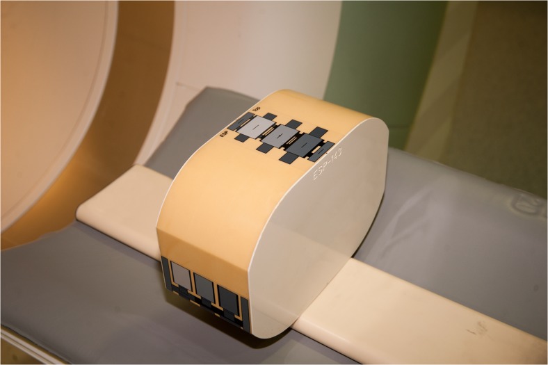
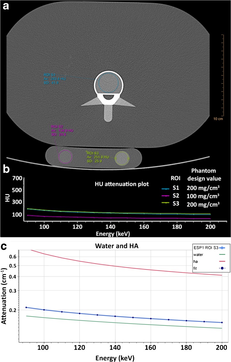
Two validated anthropomorphic phantoms containing inserts of 50-200 mg/cm calcium hydroxyapatite (HA) were scanned using a 64-slice SDCT scanner at various acquisition protocols (120 and 140 kVp, and 50, 100 and 200 mAs). Regions of interest (ROIs) were placed in each insert and mean attenuation profiles at monochromatic energy levels (90-200 keV) were constructed. These profiles were fitted to attenuation profiles of pure HA and water to calculate HA concentrations. For comparison, one phantom was scanned using dual energy X-ray absorptiometry (DXA).
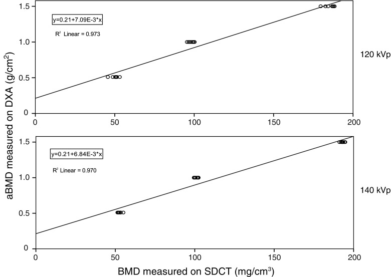
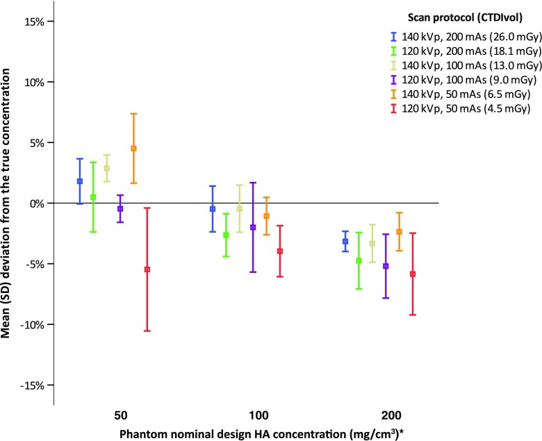
At both 120 and 140 kVp, excellent correlations (R = 0.97, P < 0.001) were found between true and measured HA concentrations. Mean error for all measurements at 120 kVp was -5.6 ± 5.7 mg/cm (-3.6 ± 3.2%) and at 140 kVp -2.4 ± 3.7 mg/cm (-0.8 ± 2.8%). Mean measurement errors were smaller than 6% for all acquisition protocols. Strong linear correlations (R ≥ 0.970, P < 0.001) with DXA were found.
SDCT allows for accurate BMD quantification and potentially opens up the possibility for osteoporosis evaluation and opportunistic screening in patients undergoing SDCT for other clinical indications. However, patient studies are needed to extend and translate our findings.
• Dual-layer spectral detector CT allows for accurate bone mineral density quantification. • BMD measurements on SDCT are strongly linearly correlated to DXA. • SDCT, acquired for several indications, may allow for evaluation of osteoporosis. • This potentially opens up the possibility for opportunistic osteoporosis screening.
研究在不同扫描方案下使用双层光谱探测器 CT(SDCT)进行骨密度(BMD)定量的准确性。
使用 64 层 SDCT 扫描仪对包含 50-200mg/cm 羟磷灰石(HA)插入物的两个经过验证的人体模型进行扫描,使用不同的采集方案(120 和 140 kVp 以及 50、100 和 200 mAs)。在每个插入物中放置感兴趣区域(ROI),并构建在单能水平(90-200keV)下的平均衰减曲线。将这些曲线拟合到纯 HA 和水的衰减曲线,以计算 HA 浓度。为了进行比较,一个模型使用双能 X 射线吸收法(DXA)进行扫描。
在 120 和 140 kVp 下,真实和测量的 HA 浓度之间均存在极好的相关性(R=0.97,P<0.001)。在 120 kVp 下,所有测量的平均误差为-5.6±5.7mg/cm(-3.6±3.2%),在 140 kVp 下为-2.4±3.7mg/cm(-0.8±2.8%)。对于所有采集方案,平均测量误差均小于 6%。与 DXA 存在很强的线性相关性(R≥0.970,P<0.001)。
SDCT 允许进行准确的 BMD 定量,并且有可能为接受 SDCT 进行其他临床适应症的患者进行骨质疏松症评估和机会性筛查开辟可能性。然而,需要进行患者研究来扩展和转化我们的发现。
双层光谱探测器 CT 允许进行准确的骨密度定量。
SDCT 上的 BMD 测量与 DXA 呈强线性相关。
用于多种适应症的 SDCT 可能允许评估骨质疏松症。
这有可能为机会性骨质疏松症筛查开辟可能性。