Nawotka K A, Huberman J A
Department of Molecular and Cellular Biology, Roswell Park Memorial Institute, Buffalo, New York 14263.
Mol Cell Biol. 1988 Apr;8(4):1408-13. doi: 10.1128/mcb.8.4.1408-1413.1988.
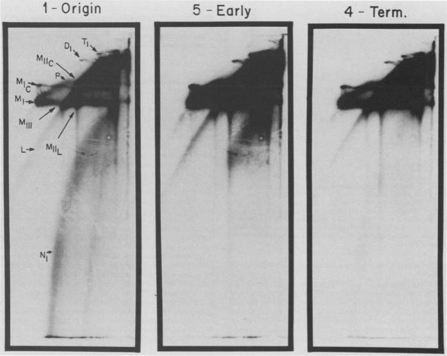
We describe in detail a method which allows determination of the directions of replication fork movement through segments of DNA for which cloned probes are available. The method uses two-dimensional neutral-alkaline agarose gel electrophoresis followed by hybridization with short probe sequences. The nascent strands of replicating molecules form an arc separated from parental and nonreplicating strands. The closer a probe is to its replication origin or to the origin-proximal end of its restriction fragment, the shorter the nascent strands that are detected by the probe. The use of multiple probes allows determination of directions of replication fork movement, as well as locations of origins and termini. In this study, we used simian virus 40 as a model to demonstrate the feasibility of the method, and we discuss its applicability to other systems.
我们详细描述了一种方法,该方法可用于确定复制叉通过可获得克隆探针的DNA片段的移动方向。该方法采用二维中性-碱性琼脂糖凝胶电泳,随后与短探针序列进行杂交。正在复制的分子的新生链形成一条与亲本链和非复制链分离的弧线。探针距离其复制起点或其限制片段的起点近端越近,该探针检测到的新生链就越短。使用多个探针可确定复制叉的移动方向,以及起点和终点的位置。在本研究中,我们以猿猴病毒40作为模型来证明该方法的可行性,并讨论其在其他系统中的适用性。