• 文献检索
  • 文档翻译
  • 深度研究
  • 学术资讯
  • Suppr Zotero 插件Zotero 插件
  • 邀请有礼
  • 套餐&价格
  • 历史记录
应用&插件
Suppr Zotero 插件Zotero 插件浏览器插件Mac 客户端Windows 客户端微信小程序
定价
高级版会员购买积分包购买API积分包
服务
文献检索文档翻译深度研究API 文档MCP 服务
关于我们
关于 Suppr公司介绍联系我们用户协议隐私条款
关注我们

Suppr 超能文献

核心技术专利:CN118964589B侵权必究
粤ICP备2023148730 号-1Suppr @ 2026

文献检索

告别复杂PubMed语法,用中文像聊天一样搜索,搜遍4000万医学文献。AI智能推荐,让科研检索更轻松。

立即免费搜索

文件翻译

保留排版,准确专业,支持PDF/Word/PPT等文件格式,支持 12+语言互译。

免费翻译文档

深度研究

AI帮你快速写综述,25分钟生成高质量综述,智能提取关键信息,辅助科研写作。

立即免费体验

用于系统微生物学的图像分析驱动的单细胞分析

Image analysis driven single-cell analytics for systems microbiology.

作者信息

Balomenos Athanasios D, Tsakanikas Panagiotis, Aspridou Zafiro, Tampakaki Anastasia P, Koutsoumanis Konstantinos P, Manolakos Elias S

机构信息

Department of Informatics and Telecommunications, National and Kapodistrian University of Athens, Ilissia, Greece.

Biomedical Research Foundation of the Academy of Athens, 4 Soranou Ephessiou Street, Athens, Greece.

出版信息

BMC Syst Biol. 2017 Apr 4;11(1):43. doi: 10.1186/s12918-017-0399-z.

DOI:10.1186/s12918-017-0399-z
PMID:28376782
原文链接:https://pmc.ncbi.nlm.nih.gov/articles/PMC5379763/
Abstract

BACKGROUND

Time-lapse microscopy is an essential tool for capturing and correlating bacterial morphology and gene expression dynamics at single-cell resolution. However state-of-the-art computational methods are limited in terms of the complexity of cell movies that they can analyze and lack of automation. The proposed Bacterial image analysis driven Single Cell Analytics (BaSCA) computational pipeline addresses these limitations thus enabling high throughput systems microbiology.

RESULTS

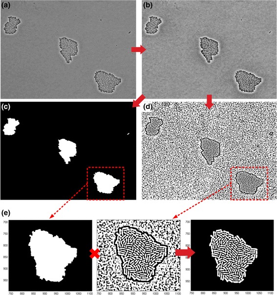
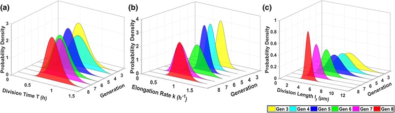
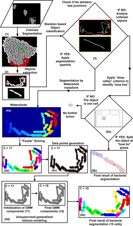
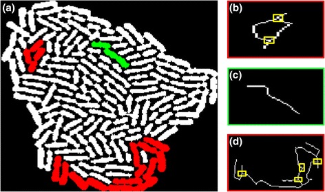
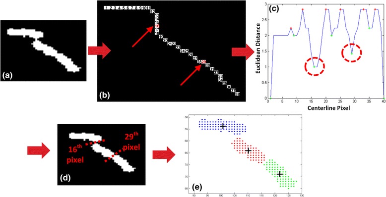
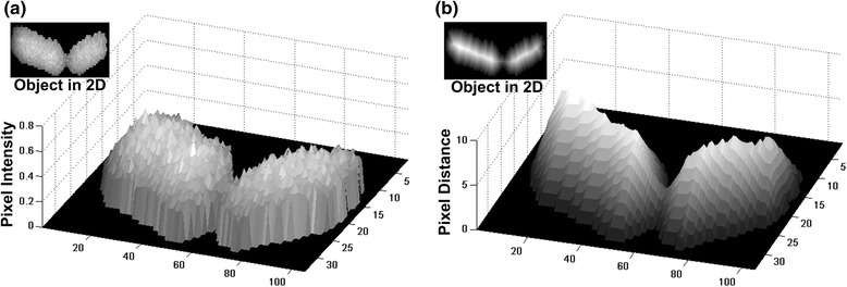
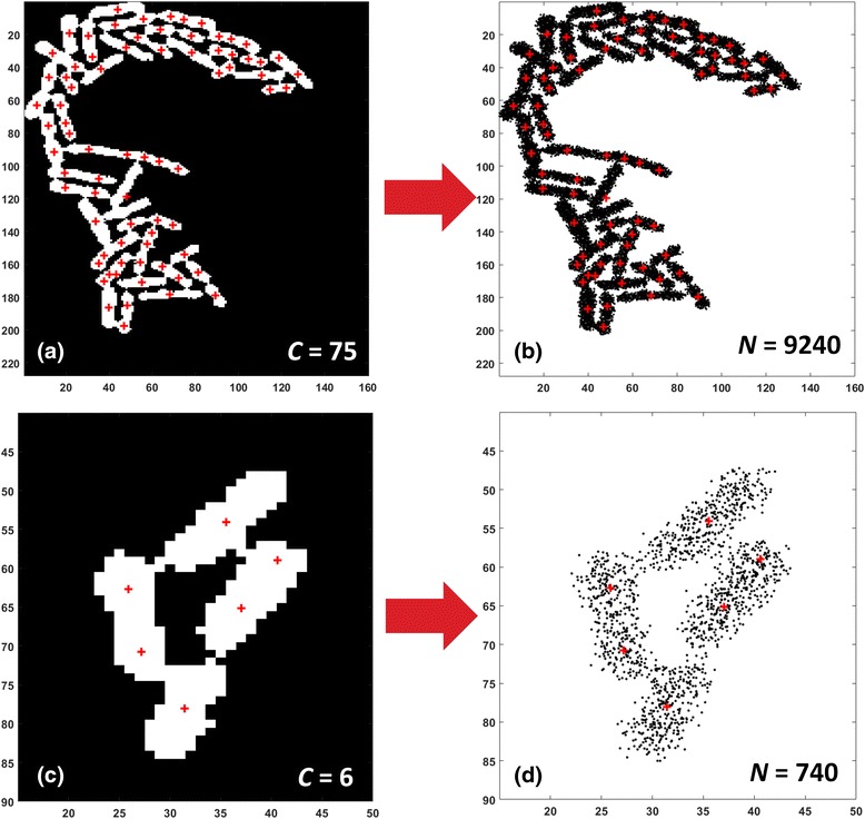
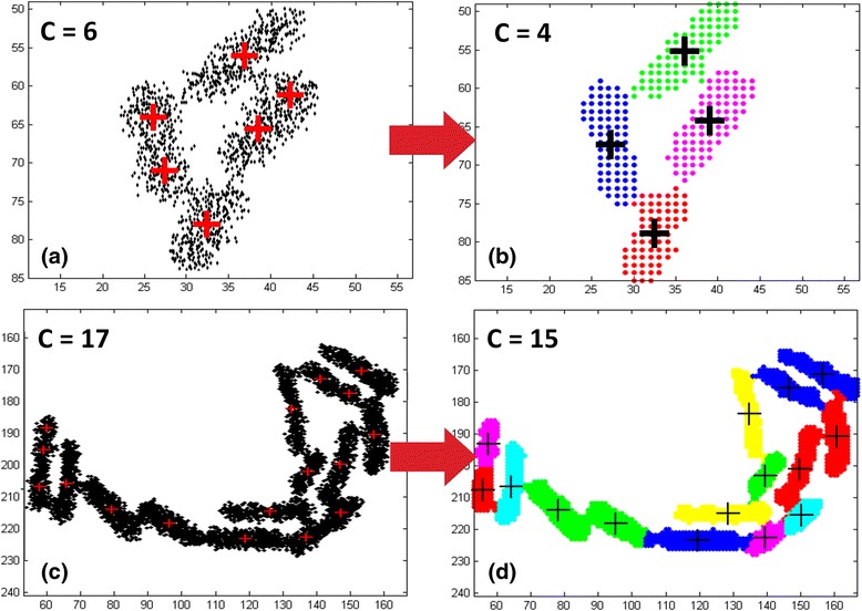
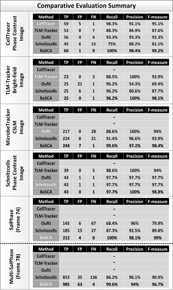
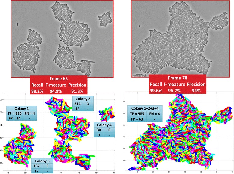
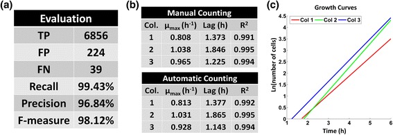
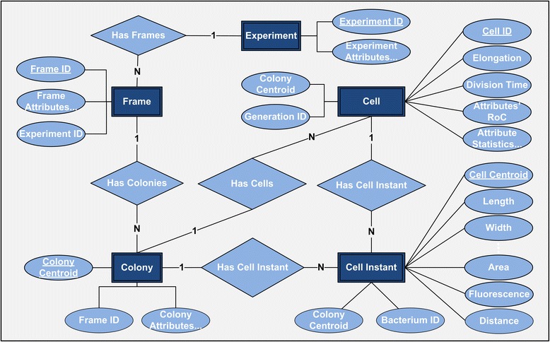
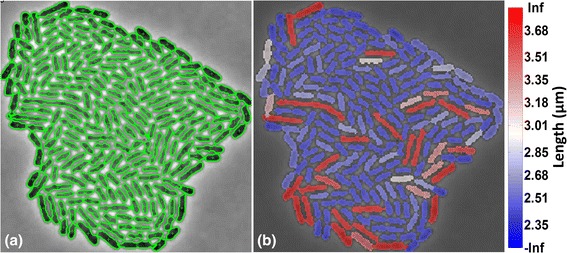
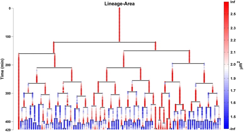
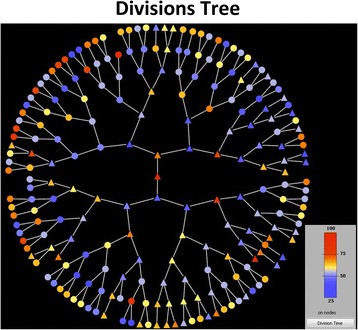
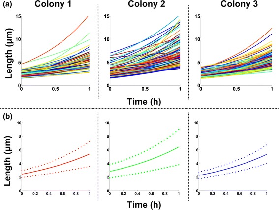
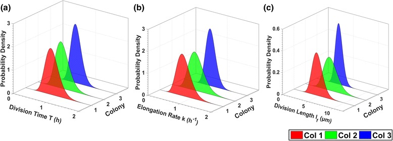
BaSCA can segment and track multiple bacterial colonies and single-cells, as they grow and divide over time (cell segmentation and lineage tree construction) to give rise to dense communities with thousands of interacting cells in the field of view. It combines advanced image processing and machine learning methods to deliver very accurate bacterial cell segmentation and tracking (F-measure over 95%) even when processing images of imperfect quality with several overcrowded colonies in the field of view. In addition, BaSCA extracts on the fly a plethora of single-cell properties, which get organized into a database summarizing the analysis of the cell movie. We present alternative ways to analyze and visually explore the spatiotemporal evolution of single-cell properties in order to understand trends and epigenetic effects across cell generations. The robustness of BaSCA is demonstrated across different imaging modalities and microscopy types.

CONCLUSIONS

BaSCA can be used to analyze accurately and efficiently cell movies both at a high resolution (single-cell level) and at a large scale (communities with many dense colonies) as needed to shed light on e.g. how bacterial community effects and epigenetic information transfer play a role on important phenomena for human health, such as biofilm formation, persisters' emergence etc. Moreover, it enables studying the role of single-cell stochasticity without losing sight of community effects that may drive it.

摘要

背景

延时显微镜是一种用于在单细胞分辨率下捕捉和关联细菌形态与基因表达动态的重要工具。然而,目前的先进计算方法在可分析的细胞电影复杂性方面存在局限性,且缺乏自动化。所提出的细菌图像分析驱动的单细胞分析(BaSCA)计算流程解决了这些局限性,从而实现了高通量系统微生物学。

结果

BaSCA能够对多个细菌菌落和单细胞进行分割和跟踪,随着它们随时间生长和分裂(细胞分割和谱系树构建),在视野中形成包含数千个相互作用细胞的密集群落。它结合了先进的图像处理和机器学习方法,即使在处理视野中存在多个过度拥挤菌落的质量不佳图像时,也能实现非常准确的细菌细胞分割和跟踪(F值超过95%)。此外,BaSCA能即时提取大量单细胞属性,并将其整理成一个数据库,总结细胞电影的分析结果。我们提出了分析和直观探索单细胞属性时空演变的替代方法,以了解细胞世代间的趋势和表观遗传效应。BaSCA的稳健性在不同成像模式和显微镜类型中得到了证明。

结论

BaSCA可根据需要用于准确高效地分析高分辨率(单细胞水平)和大规模(具有许多密集菌落的群落)的细胞电影,以阐明例如细菌群落效应和表观遗传信息传递如何在对人类健康重要的现象(如生物膜形成、持久性细胞出现等)中发挥作用。此外,它能够研究单细胞随机性的作用,而不会忽视可能驱动它的群落效应。

https://cdn.ncbi.nlm.nih.gov/pmc/blobs/ec0f/5379763/d8df8c3d1d57/12918_2017_399_Fig17_HTML.jpg
https://cdn.ncbi.nlm.nih.gov/pmc/blobs/ec0f/5379763/18862af9a8cf/12918_2017_399_Fig1_HTML.jpg
https://cdn.ncbi.nlm.nih.gov/pmc/blobs/ec0f/5379763/16a9a638418f/12918_2017_399_Fig2_HTML.jpg
https://cdn.ncbi.nlm.nih.gov/pmc/blobs/ec0f/5379763/bc30154d879a/12918_2017_399_Fig3_HTML.jpg
https://cdn.ncbi.nlm.nih.gov/pmc/blobs/ec0f/5379763/bafe8b365e0b/12918_2017_399_Fig4_HTML.jpg
https://cdn.ncbi.nlm.nih.gov/pmc/blobs/ec0f/5379763/46e0abca1460/12918_2017_399_Fig5_HTML.jpg
https://cdn.ncbi.nlm.nih.gov/pmc/blobs/ec0f/5379763/a99afe5efdbc/12918_2017_399_Fig6_HTML.jpg
https://cdn.ncbi.nlm.nih.gov/pmc/blobs/ec0f/5379763/62b43ce6ec61/12918_2017_399_Fig7_HTML.jpg
https://cdn.ncbi.nlm.nih.gov/pmc/blobs/ec0f/5379763/bced84212011/12918_2017_399_Fig8_HTML.jpg
https://cdn.ncbi.nlm.nih.gov/pmc/blobs/ec0f/5379763/5902f989f856/12918_2017_399_Fig9_HTML.jpg
https://cdn.ncbi.nlm.nih.gov/pmc/blobs/ec0f/5379763/7442c7b6aecc/12918_2017_399_Fig10_HTML.jpg
https://cdn.ncbi.nlm.nih.gov/pmc/blobs/ec0f/5379763/f5fad5381138/12918_2017_399_Fig11_HTML.jpg
https://cdn.ncbi.nlm.nih.gov/pmc/blobs/ec0f/5379763/4d04ffd47e1f/12918_2017_399_Fig12_HTML.jpg
https://cdn.ncbi.nlm.nih.gov/pmc/blobs/ec0f/5379763/79e9eec8f972/12918_2017_399_Fig13_HTML.jpg
https://cdn.ncbi.nlm.nih.gov/pmc/blobs/ec0f/5379763/0003f2d5adf4/12918_2017_399_Fig14_HTML.jpg
https://cdn.ncbi.nlm.nih.gov/pmc/blobs/ec0f/5379763/7d5fdf89c064/12918_2017_399_Fig15_HTML.jpg
https://cdn.ncbi.nlm.nih.gov/pmc/blobs/ec0f/5379763/f45ab68e1c51/12918_2017_399_Fig16_HTML.jpg
https://cdn.ncbi.nlm.nih.gov/pmc/blobs/ec0f/5379763/d8df8c3d1d57/12918_2017_399_Fig17_HTML.jpg

相似文献

1
Image analysis driven single-cell analytics for systems microbiology.用于系统微生物学的图像分析驱动的单细胞分析
BMC Syst Biol. 2017 Apr 4;11(1):43. doi: 10.1186/s12918-017-0399-z.
2
Analytics and visualization tools to characterize single-cell stochasticity using bacterial single-cell movie cytometry data.使用细菌单细胞电影细胞术数据对单细胞随机性进行特征分析和可视化工具。
BMC Bioinformatics. 2021 Oct 29;22(1):531. doi: 10.1186/s12859-021-04409-9.
3
A Cell Segmentation/Tracking Tool Based on Machine Learning.一种基于机器学习的细胞分割/追踪工具。
Methods Mol Biol. 2019;2040:399-422. doi: 10.1007/978-1-4939-9686-5_19.
4
An automatic method for robust and fast cell detection in bright field images from high-throughput microscopy.一种用于高通量显微镜明场图像中进行稳健快速细胞检测的自动方法。
BMC Bioinformatics. 2013 Oct 4;14:297. doi: 10.1186/1471-2105-14-297.
5
DeLTA: Automated cell segmentation, tracking, and lineage reconstruction using deep learning.DELTA:使用深度学习进行自动化细胞分割、跟踪和谱系重建。
PLoS Comput Biol. 2020 Apr 13;16(4):e1007673. doi: 10.1371/journal.pcbi.1007673. eCollection 2020 Apr.
6
SuperSegger: robust image segmentation, analysis and lineage tracking of bacterial cells.SuperSegger:细菌细胞的稳健图像分割、分析及谱系追踪
Mol Microbiol. 2016 Nov;102(4):690-700. doi: 10.1111/mmi.13486. Epub 2016 Sep 23.
7
Single-cell segmentation in bacterial biofilms with an optimized deep learning method enables tracking of cell lineages and measurements of growth rates.利用优化的深度学习方法对细菌生物膜中的单细胞进行分割,可实现细胞谱系追踪和生长速率测量。
Mol Microbiol. 2023 Jun;119(6):659-676. doi: 10.1111/mmi.15064. Epub 2023 Apr 17.
8
BactImAS: a platform for processing and analysis of bacterial time-lapse microscopy movies.BactImAS:用于处理和分析细菌延时显微镜电影的平台。
BMC Bioinformatics. 2014 Jul 25;15(1):251. doi: 10.1186/1471-2105-15-251.
9
Empirical gradient threshold technique for automated segmentation across image modalities and cell lines.用于跨图像模态和细胞系自动分割的经验梯度阈值技术。
J Microsc. 2015 Oct;260(1):86-99. doi: 10.1111/jmi.12269. Epub 2015 Jun 5.
10
TLM-Tracker: software for cell segmentation, tracking and lineage analysis in time-lapse microscopy movies.TLM-Tracker:用于延时显微镜电影中的细胞分割、跟踪和谱系分析的软件。
Bioinformatics. 2012 Sep 1;28(17):2276-7. doi: 10.1093/bioinformatics/bts424. Epub 2012 Jul 5.

引用本文的文献

1
Pheromone cCF10 inhibits the antibiotic persistence of by modulating energy metabolism.信息素cCF10通过调节能量代谢抑制抗生素耐受性。
Front Microbiol. 2024 Jul 8;15:1408701. doi: 10.3389/fmicb.2024.1408701. eCollection 2024.
2
Omnipose: a high-precision morphology-independent solution for bacterial cell segmentation.Omnipose:一种高精度、形态独立的细菌细胞分割解决方案。
Nat Methods. 2022 Nov;19(11):1438-1448. doi: 10.1038/s41592-022-01639-4. Epub 2022 Oct 17.
3
Segmentation, tracking and cell cycle analysis of live-cell imaging data with Cell-ACDC.

本文引用的文献

1
Noise-driven growth rate gain in clonal cellular populations.克隆细胞群体中噪声驱动的生长速率增加
Proc Natl Acad Sci U S A. 2016 Mar 22;113(12):3251-6. doi: 10.1073/pnas.1519412113. Epub 2016 Mar 7.
2
Architectural transitions in Vibrio cholerae biofilms at single-cell resolution.霍乱弧菌生物膜中单细胞分辨率下的结构转变
Proc Natl Acad Sci U S A. 2016 Apr 5;113(14):E2066-72. doi: 10.1073/pnas.1601702113. Epub 2016 Mar 1.
3
Tracking single-cells in overcrowded bacterial colonies.追踪过度拥挤细菌菌落中的单细胞。
使用 Cell-ACDC 对活细胞成像数据进行分割、跟踪和细胞周期分析。
BMC Biol. 2022 Aug 5;20(1):174. doi: 10.1186/s12915-022-01372-6.
4
Listeria monocytogenes Sublethal Injury and Viable-but-Nonculturable State Induced by Acidic Conditions and Disinfectants.酸性条件和消毒剂诱导单核细胞增生李斯特菌亚致死损伤和存活但非可培养状态。
Microbiol Spectr. 2021 Dec 22;9(3):e0137721. doi: 10.1128/Spectrum.01377-21. Epub 2021 Dec 15.
5
Analytics and visualization tools to characterize single-cell stochasticity using bacterial single-cell movie cytometry data.使用细菌单细胞电影细胞术数据对单细胞随机性进行特征分析和可视化工具。
BMC Bioinformatics. 2021 Oct 29;22(1):531. doi: 10.1186/s12859-021-04409-9.
6
Advances and opportunities in image analysis of bacterial cells and communities.细菌细胞和群落的图像分析的进展和机遇。
FEMS Microbiol Rev. 2021 Aug 17;45(4). doi: 10.1093/femsre/fuaa062.
7
Automated cell segmentation in FIJI® using the DRAQ5 nuclear dye.在 FIJI®中使用 DRAQ5 核染料进行自动细胞分割。
BMC Bioinformatics. 2019 Jan 18;20(1):39. doi: 10.1186/s12859-019-2602-2.
Annu Int Conf IEEE Eng Med Biol Soc. 2015;2015:6473-6. doi: 10.1109/EMBC.2015.7319875.
4
Oufti: an integrated software package for high-accuracy, high-throughput quantitative microscopy analysis.Oufti:一款用于高精度、高通量定量显微镜分析的集成软件包。
Mol Microbiol. 2016 Feb;99(4):767-77. doi: 10.1111/mmi.13264. Epub 2015 Dec 18.
5
Chromosomal Arrangement of Phosphorelay Genes Couples Sporulation and DNA Replication.磷中继基因的染色体排列将孢子形成与DNA复制联系起来。
Cell. 2015 Jul 16;162(2):328-337. doi: 10.1016/j.cell.2015.06.012. Epub 2015 Jul 9.
6
Scaling laws governing stochastic growth and division of single bacterial cells.支配单个细菌细胞随机生长和分裂的标度律。
Proc Natl Acad Sci U S A. 2014 Nov 11;111(45):15912-7. doi: 10.1073/pnas.1403232111. Epub 2014 Oct 27.
7
Concerted control of Escherichia coli cell division.协调控制大肠杆菌细胞分裂。
Proc Natl Acad Sci U S A. 2014 Mar 4;111(9):3431-5. doi: 10.1073/pnas.1313715111. Epub 2014 Feb 18.
8
Cell-cell contacts confine public goods diffusion inside Pseudomonas aeruginosa clonal microcolonies.细胞间接触将铜绿假单胞菌克隆微菌落中的公共物品扩散限制在内部。
Proc Natl Acad Sci U S A. 2013 Jul 30;110(31):12577-82. doi: 10.1073/pnas.1301428110. Epub 2013 Jul 15.
9
You are what you talk: quorum sensing induces individual morphologies and cell division modes in Dinoroseobacter shibae.你言我语:群体感应诱导雪双歧杆菌个体形态和细胞分裂模式。
ISME J. 2013 Dec;7(12):2274-86. doi: 10.1038/ismej.2013.107. Epub 2013 Jul 4.
10
Recruitment, assembly, and molecular architecture of the SpoIIIE DNA pump revealed by superresolution microscopy.超分辨率显微镜揭示 SpoIIIE DNA 泵的招募、组装和分子结构。
PLoS Biol. 2013;11(5):e1001557. doi: 10.1371/journal.pbio.1001557. Epub 2013 May 7.