Department of Cell Biology, The Johns Hopkins University School of Medicine, 725N. Wolfe St., Baltimore, MD, 21205, USA.
The Johns Hopkins Malaria Research Institute, The Johns Hopkins University School of Medicine, 725N. Wolfe St., Baltimore, MD, 21205, USA.
Sci Rep. 2017 Apr 4;7(1):601. doi: 10.1038/s41598-017-00672-0.
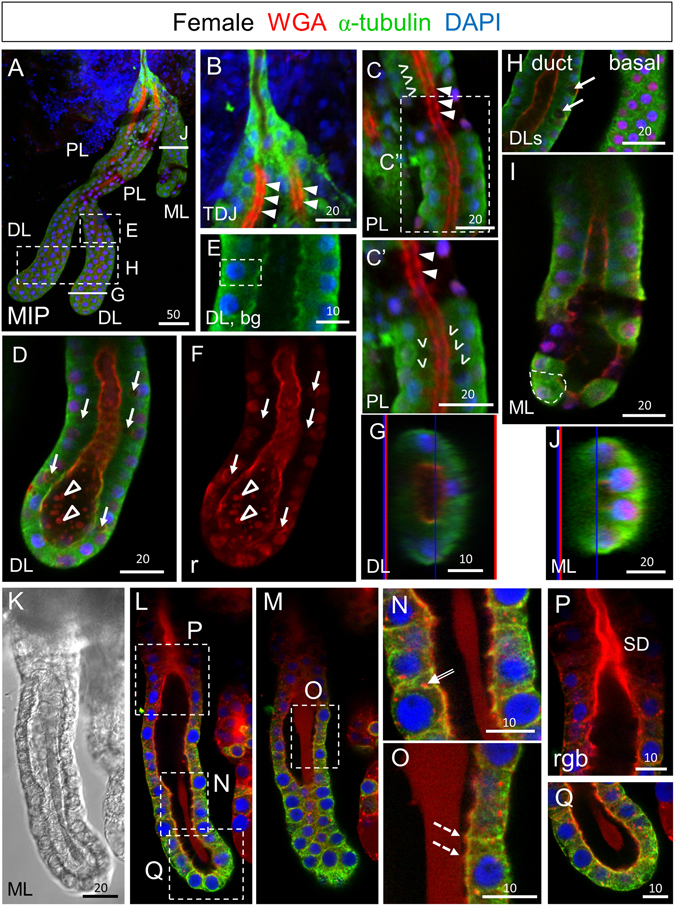
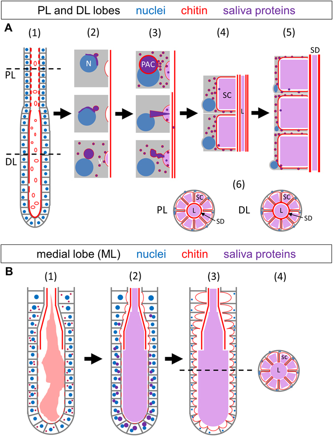
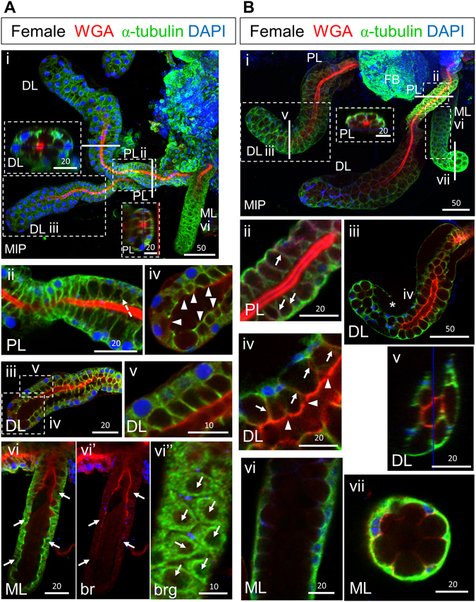
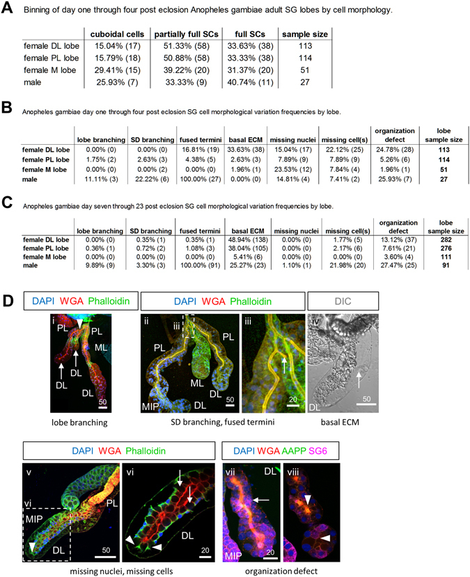
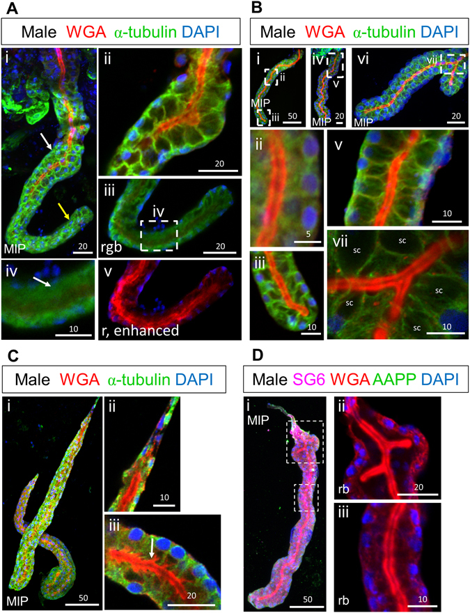
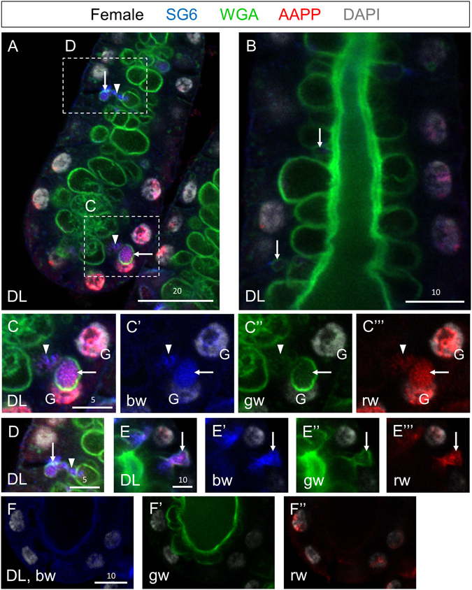
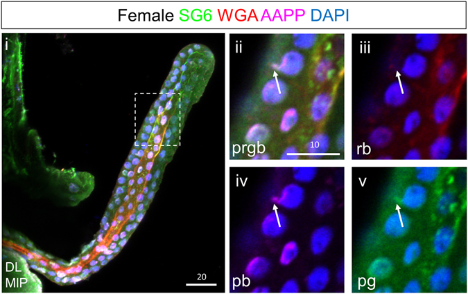
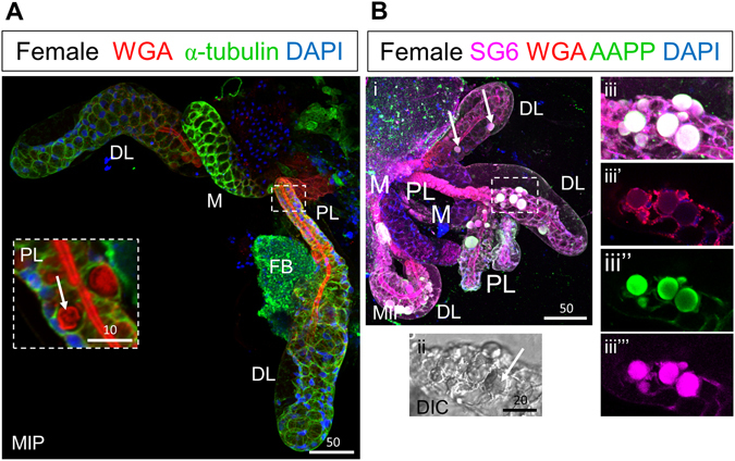
Mosquito-borne diseases cause one million deaths and hundreds of millions of human infections yearly. With all such diseases, the pathogen must traverse the mosquito salivary gland (SG) for transmission to a new host, making the SGs ideal targets for genetic strategies to block transmission. Prior studies have elucidated details of SG structure by light and electron microscopy and have deeply explored the salivary transcriptome and proteome. Very little is known, however, about how the unique functional architecture of mosquito SGs is achieved. Using immunohistochemistry and confocal microscopy, we address two questions regarding SGs of the malaria vector Anopheles gambiae. How does the distinct cup-shaped morphology of SG secretory cells arise? And, how does the salivary duct, the structure through which saliva and parasites exit the glands, form? We demonstrate that SG cells begin as cuboidal-shaped cells surrounding a matrix-filled lumen that mature into cup-shaped cells through the formation and fusion of a large pre-apical compartment (PAC) to the apical surface. The secretory duct begins as buds of chitin at the apical surface of individual secretory cells. Further chitin deposition connects these chitin buds to form a contiguous duct that largely separates from the apical surface during PAC fusion.
蚊媒疾病每年导致 100 万人死亡和数亿人感染。对于所有此类疾病,病原体必须穿过蚊子的唾液腺 (SG) 才能传播给新宿主,因此 SG 是阻断传播的遗传策略的理想目标。先前的研究通过光镜和电子显微镜阐明了 SG 结构的细节,并深入研究了唾液转录组和蛋白质组。然而,关于蚊子 SG 的独特功能结构是如何实现的,我们知之甚少。使用免疫组织化学和共聚焦显微镜,我们解决了有关疟疾病媒按蚊 SG 的两个问题。SG 分泌细胞独特的杯状形态是如何产生的?以及,唾液和寄生虫离开腺体的唾液道是如何形成的?我们证明,SG 细胞最初是围绕充满基质的腔的立方体形细胞,通过大的前尖室 (PAC) 形成和融合到顶表面,成熟为杯状细胞。分泌道最初是单个分泌细胞顶表面的几丁质芽。进一步的几丁质沉积将这些几丁质芽连接起来,形成一个连续的管,在 PAC 融合期间大部分与顶表面分离。