Department of Cell Biology, Johns Hopkins School of Medicine, Baltimore, Maryland, USA.
Johns Hopkins Malaria Research Institute, Johns Hopkins Bloomberg School of Public Health, Baltimore, Maryland, USA.
mBio. 2019 Aug 6;10(4):e01238-19. doi: 10.1128/mBio.01238-19.
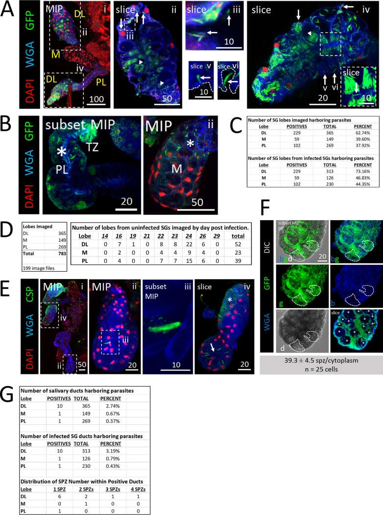
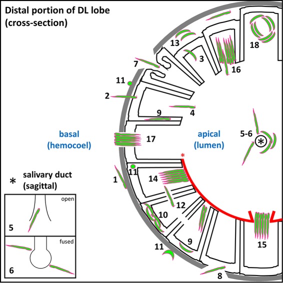
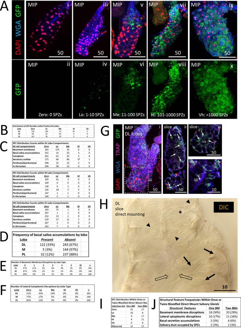
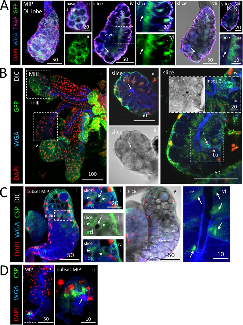
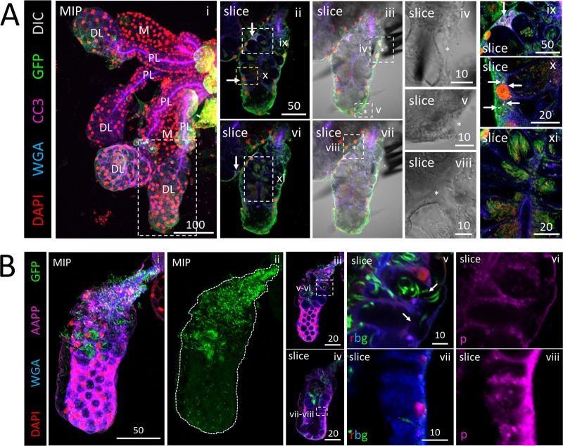
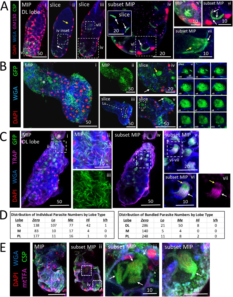
sporozoites (SPZs) must traverse the mosquito salivary glands (SGs) to reach a new vertebrate host and continue the malaria disease cycle. Although SGs can harbor thousands of sporozoites, only 10 to 100 are deposited into a host during probing. To determine how the SGs might function as a bottleneck in SPZ transmission, we have characterized SGs infected with the rodent malaria parasite using immunofluorescence confocal microscopy. Our analyses corroborate findings from previous electron microscopy studies and provide new insights into the invasion process. We identified sites of SPZ accumulation within SGs across a range of infection intensities. Although SPZs were most often seen in the distal lateral SG lobes, they were also observed in the medial and proximal lateral lobes. Most parasites were associated with either the basement membrane or secretory cavities. SPZs accumulated at physical barriers, including fused salivary ducts and extensions of the chitinous salivary duct wall into the distal lumen. SPZs were observed only rarely within salivary ducts. SPZs appeared to contact each other in many different quantities, not just in the previously described large bundles. Within parasite bundles, all of the SPZs were oriented in the same direction. We found that moderate levels of infection did not necessarily correlate with major SG disruptions or abundant SG cell death. Altogether, our findings suggest that SG architecture largely acts as a barrier to SPZ transmission. Malaria continues to have a devastating impact on human health. With growing resistance to insecticides and antimalarial drugs, as well as climate change predictions indicating expansion of vector territories, the impact of malaria is likely to increase. Additional insights regarding pathogen migration through vector mosquitoes are needed to develop novel methods to prevent transmission to new hosts. Pathogens, including the microbes that cause malaria, must invade the salivary glands (SGs) for transmission. Since SG traversal is required for parasite transmission, SGs are ideal targets for transmission-blocking strategies. The work presented here highlights the role that mosquito SG architecture plays in limiting parasite traversal, revealing how the SG transmission bottleneck is imposed. Further, our data provide unprecedented detail about SG-sporozoite interactions and gland-to-gland variation not provided in previous studies.
疟原虫的子孢子(SPZ)必须穿过蚊子的唾液腺(SG)才能到达新的脊椎动物宿主并继续疟疾的疾病循环。尽管 SG 可以容纳数千个子孢子,但在探测过程中只有 10 到 100 个子孢子被沉积到宿主中。为了确定 SG 如何作为 SPZ 传播的瓶颈,我们使用免疫荧光共聚焦显微镜对感染啮齿动物疟原虫的 SG 进行了特征描述。我们的分析证实了以前电子显微镜研究的结果,并为入侵过程提供了新的见解。我们在一系列感染强度范围内确定了 SG 中 SPZ 积累的部位。尽管 SPZ 最常出现在 SG 的远端外侧叶,但也出现在内侧和近端外侧叶。大多数寄生虫与基膜或分泌腔有关。SPZ 在物理屏障处积累,包括融合的唾液管和几丁质唾液管壁向远端管腔的延伸。很少观察到 SPZ 在唾液管内。SPZ 似乎以多种不同的数量相互接触,而不仅仅是以前描述的大束。在寄生虫束内,所有的 SPZ 都朝同一方向定向。我们发现,中度感染水平不一定与 SG 严重破坏或大量 SG 细胞死亡相关。总的来说,我们的研究结果表明,SG 结构在很大程度上充当了 SPZ 传播的屏障。疟疾继续对人类健康造成毁灭性影响。随着杀虫剂和抗疟药物耐药性的增加,以及气候变化预测表明媒介领土的扩大,疟疾的影响可能会增加。需要进一步了解病原体通过媒介蚊子的迁移,以开发新的方法来防止传播给新的宿主。病原体,包括引起疟疾的微生物,必须侵入唾液腺(SG)才能传播。由于 SG 的穿透是寄生虫传播所必需的,因此 SG 是阻断传播策略的理想目标。这里介绍的工作强调了蚊子 SG 结构在限制寄生虫穿透方面的作用,揭示了 SG 传播瓶颈是如何产生的。此外,我们的数据提供了以前研究中未提供的关于 SG-子孢子相互作用和腺体之间差异的前所未有的详细信息。