• 文献检索
  • 文档翻译
  • 深度研究
  • 学术资讯
  • Suppr Zotero 插件Zotero 插件
  • 邀请有礼
  • 套餐&价格
  • 历史记录
应用&插件
Suppr Zotero 插件Zotero 插件浏览器插件Mac 客户端Windows 客户端微信小程序
定价
高级版会员购买积分包购买API积分包
服务
文献检索文档翻译深度研究API 文档MCP 服务
关于我们
关于 Suppr公司介绍联系我们用户协议隐私条款
关注我们

Suppr 超能文献

核心技术专利:CN118964589B侵权必究
粤ICP备2023148730 号-1Suppr @ 2026

文献检索

告别复杂PubMed语法,用中文像聊天一样搜索,搜遍4000万医学文献。AI智能推荐,让科研检索更轻松。

立即免费搜索

文件翻译

保留排版,准确专业,支持PDF/Word/PPT等文件格式,支持 12+语言互译。

免费翻译文档

深度研究

AI帮你快速写综述,25分钟生成高质量综述,智能提取关键信息,辅助科研写作。

立即免费体验

一条Wnt平面极性信号通路通过内体信号传导限制F-肌动蛋白组装,从而指导神经突分支。

A Wnt-planar polarity pathway instructs neurite branching by restricting F-actin assembly through endosomal signaling.

作者信息

Chen Chun-Hao, He Chun-Wei, Liao Chien-Po, Pan Chun-Liang

机构信息

Institute of Molecular Medicine, College of Medicine, National Taiwan University, Taipei, Taiwan.

出版信息

PLoS Genet. 2017 Apr 6;13(4):e1006720. doi: 10.1371/journal.pgen.1006720. eCollection 2017 Apr.

DOI:10.1371/journal.pgen.1006720
PMID:28384160
原文链接:https://pmc.ncbi.nlm.nih.gov/articles/PMC5398721/
Abstract

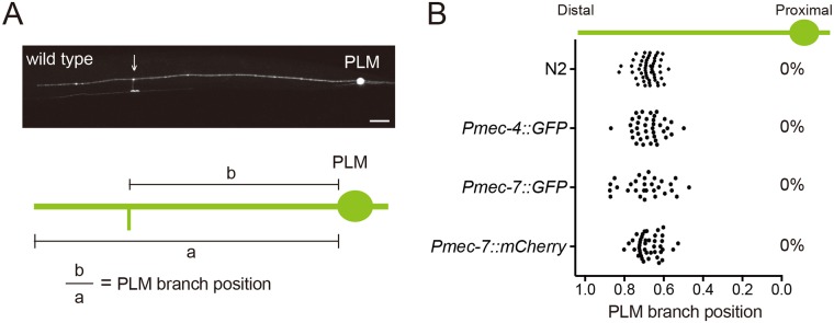
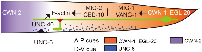
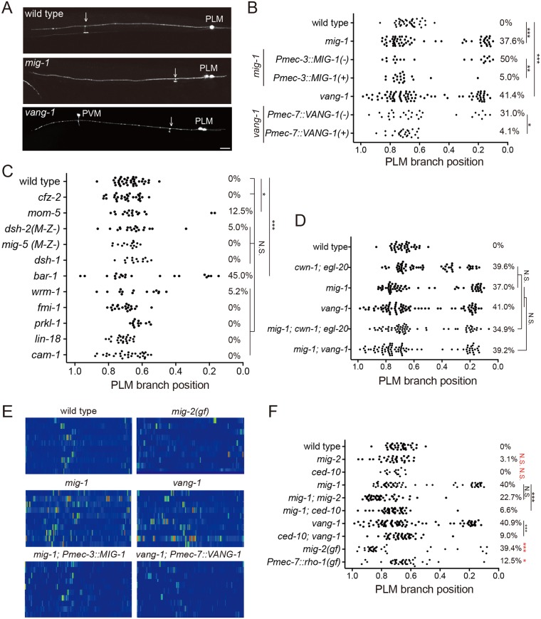
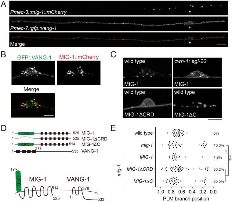
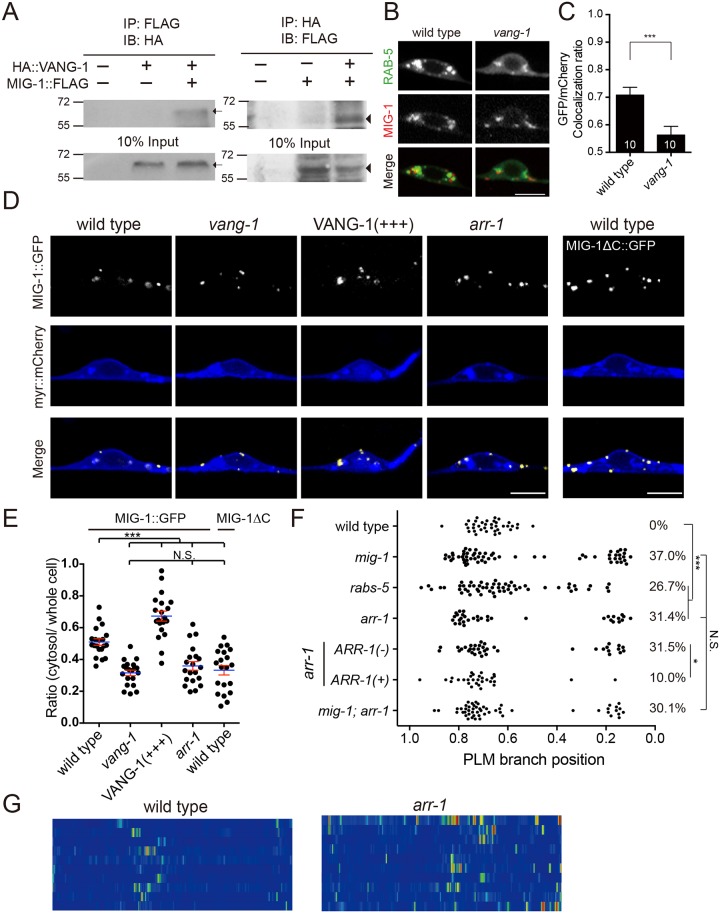
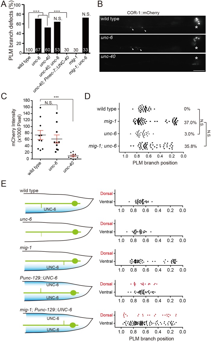
Spatial arrangement of neurite branching is instructed by both attractive and repulsive cues. Here we show that in C. elegans, the Wnt family of secreted glycoproteins specify neurite branching sites in the PLM mechanosensory neurons. Wnts function through MIG-1/Frizzled and the planar cell polarity protein (PCP) VANG-1/Strabismus/Vangl2 to restrict the formation of F-actin patches, which mark branching sites in nascent neurites. We find that VANG-1 promotes Wnt signaling by facilitating Frizzled endocytosis and genetically acts in a common pathway with arr-1/β-arrestin, whose mutation results in defective PLM branching and F-actin patterns similar to those in the Wnt, mig-1 or vang-1 mutants. On the other hand, the UNC-6/Netrin pathway intersects orthogonally with Wnt-PCP signaling to guide PLM branch growth along the dorsal-ventral axis. Our study provides insights for how attractive and repulsive signals coordinate to sculpt neurite branching patterns, which are critical for circuit connectivity.

摘要

神经突分支的空间排列受吸引和排斥信号的共同指导。我们在此表明,在秀丽隐杆线虫中,分泌型糖蛋白的Wnt家族决定了PLM机械感觉神经元中的神经突分支位点。Wnts通过MIG-1/卷曲蛋白和平面细胞极性蛋白(PCP)VANG-1/斜视蛋白/Vangl2发挥作用,以限制F-肌动蛋白斑块的形成,这些斑块标志着新生神经突中的分支位点。我们发现,VANG-1通过促进卷曲蛋白的内吞作用来促进Wnt信号传导,并且在与arr-1/β-抑制蛋白的共同途径中发挥遗传作用,其突变导致PLM分支和F-肌动蛋白模式缺陷,类似于Wnt、mig-1或vang-1突变体中的情况。另一方面,UNC-6/网蛋白途径与Wnt-PCP信号正交相交,以指导PLM分支沿背腹轴生长。我们的研究为吸引和排斥信号如何协同塑造对回路连接至关重要的神经突分支模式提供了见解。

https://cdn.ncbi.nlm.nih.gov/pmc/blobs/cfe0/5398721/74cffea16ade/pgen.1006720.g008.jpg
https://cdn.ncbi.nlm.nih.gov/pmc/blobs/cfe0/5398721/bea56a236715/pgen.1006720.g001.jpg
https://cdn.ncbi.nlm.nih.gov/pmc/blobs/cfe0/5398721/622b86992c1f/pgen.1006720.g002.jpg
https://cdn.ncbi.nlm.nih.gov/pmc/blobs/cfe0/5398721/d0e29e6af17c/pgen.1006720.g003.jpg
https://cdn.ncbi.nlm.nih.gov/pmc/blobs/cfe0/5398721/c7ce80a35fd8/pgen.1006720.g004.jpg
https://cdn.ncbi.nlm.nih.gov/pmc/blobs/cfe0/5398721/a425775b7fd6/pgen.1006720.g005.jpg
https://cdn.ncbi.nlm.nih.gov/pmc/blobs/cfe0/5398721/821dc5f8af9a/pgen.1006720.g006.jpg
https://cdn.ncbi.nlm.nih.gov/pmc/blobs/cfe0/5398721/bdbeb778d363/pgen.1006720.g007.jpg
https://cdn.ncbi.nlm.nih.gov/pmc/blobs/cfe0/5398721/74cffea16ade/pgen.1006720.g008.jpg

相似文献

1
A Wnt-planar polarity pathway instructs neurite branching by restricting F-actin assembly through endosomal signaling.一条Wnt平面极性信号通路通过内体信号传导限制F-肌动蛋白组装,从而指导神经突分支。
PLoS Genet. 2017 Apr 6;13(4):e1006720. doi: 10.1371/journal.pgen.1006720. eCollection 2017 Apr.
2
The polarity protein VANG-1 antagonizes Wnt signaling by facilitating Frizzled endocytosis.极性蛋白 VANG-1 通过促进 Frizzled 内化来拮抗 Wnt 信号。
Development. 2018 Dec 17;145(24):dev168666. doi: 10.1242/dev.168666.
3
VANG-1 and PRKL-1 cooperate to negatively regulate neurite formation in Caenorhabditis elegans.VANG-1 和 PRKL-1 合作负调控秀丽隐杆线虫的轴突形成。
PLoS Genet. 2011 Sep;7(9):e1002257. doi: 10.1371/journal.pgen.1002257. Epub 2011 Sep 1.
4
Regulation of Axon Guidance by the Wnt Receptor Ror/CAM-1 in the PVT Guidepost Cell in .Wnt 受体 Ror/CAM-1 在 PVT 导位细胞中对轴突导向的调控。
Genetics. 2017 Dec;207(4):1533-1545. doi: 10.1534/genetics.117.300375. Epub 2017 Oct 9.
5
Netrins and Wnts function redundantly to regulate antero-posterior and dorso-ventral guidance in C. elegans.在秀丽隐杆线虫中,Netrins和Wnts发挥冗余功能来调节前后和背腹导向。
PLoS Genet. 2014 Jun 5;10(6):e1004381. doi: 10.1371/journal.pgen.1004381. eCollection 2014 Jun.
6
The planar cell polarity protein VANG-1/Vangl negatively regulates Wnt/β-catenin signaling through a Dvl dependent mechanism.平面细胞极性蛋白 VANG-1/Vangl 通过 Dvl 依赖的机制负调控 Wnt/β-连环蛋白信号通路。
PLoS Genet. 2018 Dec 7;14(12):e1007840. doi: 10.1371/journal.pgen.1007840. eCollection 2018 Dec.
7
A Wnt-Frz/Ror-Dsh pathway regulates neurite outgrowth in Caenorhabditis elegans.Wnt-Frz/Ror-Dsh 通路调控秀丽隐杆线虫的神经突生长。
PLoS Genet. 2010 Aug 12;6(8):e1001056. doi: 10.1371/journal.pgen.1001056.
8
Wnt signals and frizzled activity orient anterior-posterior axon outgrowth in C. elegans.Wnt信号和卷曲蛋白活性决定了秀丽隐杆线虫前后轴突的生长方向。
Dev Cell. 2006 Mar;10(3):379-90. doi: 10.1016/j.devcel.2006.01.013.
9
Dishevelled attenuates the repelling activity of Wnt signaling during neurite outgrowth in Caenorhabditis elegans.在秀丽隐杆线虫的轴突生长过程中,蓬乱蛋白减弱Wnt信号通路的排斥活性。
Proc Natl Acad Sci U S A. 2015 Oct 27;112(43):13243-8. doi: 10.1073/pnas.1518686112. Epub 2015 Oct 12.
10
C. elegans fmi-1/flamingo and Wnt pathway components interact genetically to control the anteroposterior neurite growth of the VD GABAergic neurons.秀丽隐杆线虫 fmi-1/flamingo 和 Wnt 通路成分在遗传上相互作用,以控制 VD GABAergic 神经元的前后神经突生长。
Dev Biol. 2013 May 1;377(1):224-35. doi: 10.1016/j.ydbio.2013.01.014. Epub 2013 Jan 30.

引用本文的文献

1
Axonal mitochondria regulate gentle touch response through control of axonal actin dynamics.轴突线粒体通过控制轴突肌动蛋白动力学来调节轻触觉反应。
bioRxiv. 2024 Aug 13:2024.08.13.607780. doi: 10.1101/2024.08.13.607780.
2
β-arrestins Are Scaffolding Proteins Required for Shh-Mediated Axon Guidance.β-arrestins 作为衔接蛋白在 Shh 介导的轴突导向中发挥作用。
J Neurosci. 2024 Jul 24;44(30):e0261242024. doi: 10.1523/JNEUROSCI.0261-24.2024.
3
Wnt-Ror-Dvl signalling and the dystrophin complex organize planar-polarized membrane compartments in C. elegans muscles.

本文引用的文献

1
Specification of Dendritogenesis Site in Drosophila aCC Motoneuron by Membrane Enrichment of Pak1 through Dscam1.通过Dscam1使Pak1在膜上富集从而确定果蝇aCC运动神经元中树突发生的位点
Dev Cell. 2015 Oct 12;35(1):93-106. doi: 10.1016/j.devcel.2015.09.007.
2
Dishevelled attenuates the repelling activity of Wnt signaling during neurite outgrowth in Caenorhabditis elegans.在秀丽隐杆线虫的轴突生长过程中,蓬乱蛋白减弱Wnt信号通路的排斥活性。
Proc Natl Acad Sci U S A. 2015 Oct 27;112(43):13243-8. doi: 10.1073/pnas.1518686112. Epub 2015 Oct 12.
3
Axon Dynamics during Neocortical Laminar Innervation.
Wnt-Ror-Dvl 信号通路和肌营养不良蛋白复合物在秀丽隐杆线虫肌肉中组织平面极化的膜隔室。
Nat Commun. 2024 Jun 10;15(1):4935. doi: 10.1038/s41467-024-49154-8.
4
The Axonal Actin Filament Cytoskeleton: Structure, Function, and Relevance to Injury and Degeneration.轴突肌动蛋白丝细胞骨架:结构、功能及与损伤和变性的关系。
Mol Neurobiol. 2024 Aug;61(8):5646-5664. doi: 10.1007/s12035-023-03879-7. Epub 2024 Jan 13.
5
Synaptic branch stability is mediated by non-enzymatic functions of MEC-17/αTAT1 and ATAT-2.突触分支稳定性由 MEC-17/αTAT1 和 ATAT-2 的非酶功能介导。
Sci Rep. 2022 Aug 17;12(1):14003. doi: 10.1038/s41598-022-18333-2.
6
Transparent Touch: Insights From Model Systems on Epidermal Control of Somatosensory Innervation.透明触摸:来自模型系统对躯体感觉神经支配的表皮控制的见解。
Front Cell Neurosci. 2021 May 31;15:680345. doi: 10.3389/fncel.2021.680345. eCollection 2021.
7
Regulation of UNC-40/DCC and UNC-6/Netrin by DAF-16 promotes functional rewiring of the injured axon.UNC-40/DCC 和 UNC-6/Netrin 的调节通过 DAF-16 促进损伤轴突的功能重布线。
Development. 2021 Jun 1;148(11). doi: 10.1242/dev.198044. Epub 2021 Jun 10.
8
Extrinsic Factors Regulating Dendritic Patterning.调节树突形态形成的外在因素。
Front Cell Neurosci. 2021 Jan 13;14:622808. doi: 10.3389/fncel.2020.622808. eCollection 2020.
9
Neurite Branching Regulated by Neuronal Cell Surface Molecules in .神经元细胞表面分子对神经突分支的调控 于……
Front Neuroanat. 2020 Aug 21;14:59. doi: 10.3389/fnana.2020.00059. eCollection 2020.
10
LRRK2 mediates axon development by regulating Frizzled3 phosphorylation and growth cone-growth cone communication.LRRK2 通过调节 Frizzled3 的磷酸化和生长锥-生长锥通讯来介导轴突发育。
Proc Natl Acad Sci U S A. 2020 Jul 28;117(30):18037-18048. doi: 10.1073/pnas.1921878117. Epub 2020 Jul 8.
新皮层分层神经支配过程中的轴突动力学
Cell Rep. 2015 Jul 14;12(2):172-82. doi: 10.1016/j.celrep.2015.06.026. Epub 2015 Jul 2.
4
RHGF-1/PDZ-RhoGEF and retrograde DLK-1 signaling drive neuronal remodeling on microtubule disassembly.RHGF-1/PDZ-RhoGEF和逆行DLK-1信号通路驱动微管解聚时的神经元重塑。
Proc Natl Acad Sci U S A. 2014 Nov 18;111(46):16568-73. doi: 10.1073/pnas.1410263111. Epub 2014 Oct 30.
5
Involvement of Rho-family GTPases in axon branching.Rho家族小GTP酶在轴突分支中的作用。
Small GTPases. 2014;5:e27974. doi: 10.4161/sgtp.27974. Epub 2014 Mar 11.
6
Local F-actin network links synapse formation and axon branching.局部 F-肌动蛋白网络连接突触形成和轴突分支。
Cell. 2014 Jan 16;156(1-2):208-20. doi: 10.1016/j.cell.2013.12.009.
7
The WAVE regulatory complex links diverse receptors to the actin cytoskeleton.WAVE 调节复合物将各种受体连接到肌动蛋白细胞骨架。
Cell. 2014 Jan 16;156(1-2):195-207. doi: 10.1016/j.cell.2013.11.048.
8
The conserved transmembrane RING finger protein PLR-1 downregulates Wnt signaling by reducing Frizzled, Ror and Ryk cell-surface levels in C. elegans.保守的跨膜环指蛋白 PLR-1 通过降低秀丽隐杆线虫中 Frizzled、Ror 和 Ryk 的细胞表面水平来下调 Wnt 信号。
Development. 2014 Feb;141(3):617-28. doi: 10.1242/dev.101600. Epub 2014 Jan 8.
9
Antagonistic functions of Dishevelleds regulate Frizzled3 endocytosis via filopodia tips in Wnt-mediated growth cone guidance.Dishevelleds 的拮抗功能通过丝状伪足尖端调节 Wnt 介导的生长锥导向中的 Frizzled3 内吞作用。
J Neurosci. 2013 Dec 4;33(49):19071-85. doi: 10.1523/JNEUROSCI.2800-13.2013.
10
Two Wnts instruct topographic synaptic innervation in C. elegans.两种Wnt蛋白指导秀丽隐杆线虫的拓扑突触神经支配。
Cell Rep. 2013 Oct 31;5(2):389-96. doi: 10.1016/j.celrep.2013.09.011. Epub 2013 Oct 17.