Anders Fabian, Teister Julia, Liu Aiwei, Funke Sebastian, Grus Franz H, Thanos Solon, von Pein Harald D, Pfeiffer Norbert, Prokosch Verena
Experimental and Translational Ophthalmology, Department of Ophthalmology, University Medical Center of the Johannes Gutenberg-University Mainz, Mainz, Germany.
Institute for Experimental Ophthalmology, School of Medicine, Westfalian-Wilhelms-University Münster, Münster, Germany.
PLoS One. 2017 Apr 6;12(4):e0175451. doi: 10.1371/journal.pone.0175451. eCollection 2017.
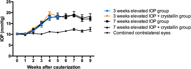
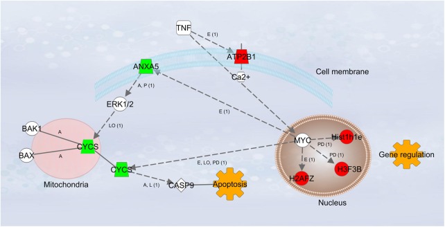
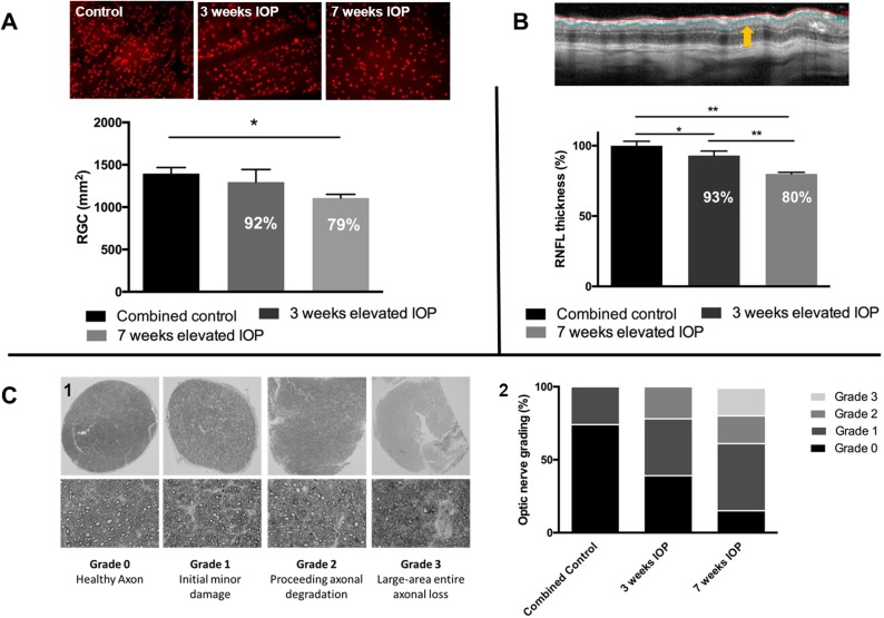
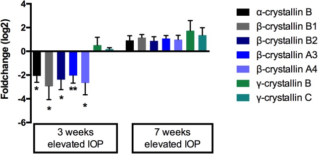
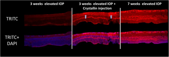
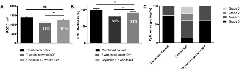
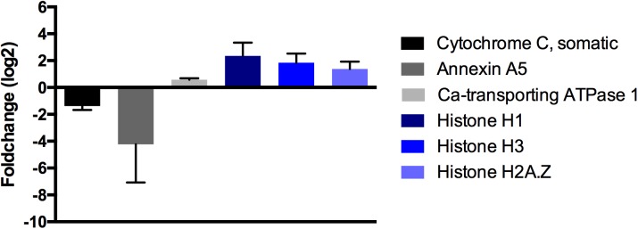
Purpose of this study was to investigate firstly specific proteomic changes within the retina in the course of an animal glaucoma model and to identify secondly new approaches for neuroprotective, therapeutic options in glaucoma by addressing those specific changes. Intraocular pressure was elevated through cauterization of episcleral veins in adult Sprague Dawley rats. Molecular and morphological changes were surveyed using mass spectrometry, optical coherence tomography as well as immunohistochemical cross section- and flat mount stainings. By quantifying more than 1500 retinal proteins, it was found that the HspB5 protein and numerous beta-crystallins showed a uniform and unique shifting expression pattern as a result of different periods of elevated IOP exposure. Crystallins showed a significant downregulation (p<0.05) after 3 weeks of elevated IOP and an upregulation after 7 weeks. Counteracting those typical changes, an intravitreal injection of β-crystallin B2 at the time of IOP elevation was found to reduce retinal ganglion cell loss (p<0.05), decrease of the retinal nerve fiber layer (p<0.05) and impairment of the optic nerve. Ultimately, proteomic data revealed that β-crystallin B2 might influence calcium-depended cell signaling pathways with severe effect on apoptosis and gene regulation. In this context especially annexin A5, calcium-transporting ATPase 1 and various histone proteins seem to play a major role.
本研究的目的首先是调查动物青光眼模型过程中视网膜内特定的蛋白质组学变化,其次是通过解决这些特定变化来确定青光眼神经保护治疗选择的新方法。通过烧灼成年Sprague Dawley大鼠的巩膜静脉来升高眼压。使用质谱、光学相干断层扫描以及免疫组织化学横断面和整装片染色来调查分子和形态学变化。通过对1500多种视网膜蛋白进行定量分析,发现由于不同时间段的眼压升高暴露,HspB5蛋白和许多β-晶状体蛋白呈现出一致且独特的表达模式变化。晶状体蛋白在眼压升高3周后显著下调(p<0.05),7周后上调。为抵消这些典型变化,发现在眼压升高时玻璃体腔内注射β-晶状体蛋白B2可减少视网膜神经节细胞丢失(p<0.05)、视网膜神经纤维层厚度降低(p<严重影响凋亡和基因调控。在此背景下,尤其是膜联蛋白A5、钙转运ATP酶1和各种组蛋白似乎起主要作用。 05)以及视神经损伤。最终,蛋白质组学数据表明β-晶状体蛋白B2可能影响依赖钙的细胞信号通路,对凋亡和基因调控产生严重影响。在此背景下,尤其是膜联蛋白A5、钙转运ATP酶1和各种组蛋白似乎起主要作用。