Uitterdijk André, Springeling Tirza, Hermans Kevin C M, Merkus Daphne, de Beer Vincent J, Gorsse-Bakker Charlotte, Mokelke Eric, Daskalopoulos Evangelos P, Wielopolski Piotr A, Cleutjens Jack P M, Blankesteijn W Matthijs, Prinzen Frits W, van der Giessen Willem J, van Geuns Robert-Jan M, Duncker Dirk J
Department of Cardiology, Ee-2351, Erasmus MC, University Medical Center Rotterdam, PO Box 2040, 3000 CA, Rotterdam, The Netherlands.
Department of Radiology, Erasmus MC, Rotterdam, The Netherlands.
Basic Res Cardiol. 2017 May;112(3):28. doi: 10.1007/s00395-017-0616-3. Epub 2017 Apr 6.
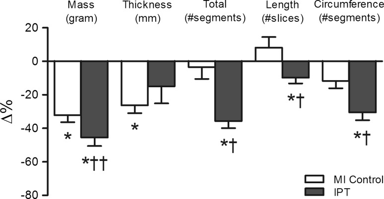
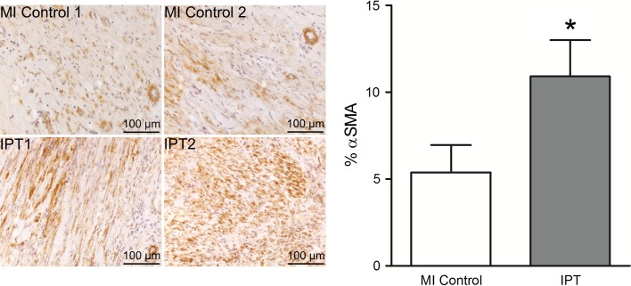
Despite early revascularization, remodeling and dysfunction of the left ventricle (LV) after acute myocardial infarction (AMI) remain important therapeutic targets. Intermittent pacing therapy (IPT) of the LV can limit infarct size, when applied during early reperfusion. However, the effects of IPT on post-AMI LV remodeling and infarct healing are unknown. We therefore investigated the effects of IPT on global LV remodeling and infarct geometry in swine with a 3-day old AMI. For this purpose, fifteen pigs underwent 2 h ligation of the left circumflex coronary artery followed by reperfusion. An epicardial pacing lead was implanted in the peri-infarct zone. After three days, global LV remodeling and infarct geometry were assessed using magnetic resonance imaging (MRI). Animals were stratified into MI control and IPT groups. Thirty-five days post-AMI, follow-up MRI was obtained and myofibroblast content, markers of extracellular matrix (ECM) turnover and Wnt/frizzled signaling in infarct and non-infarct control tissue were studied. Results showed that IPT had no significant effect on global LV remodeling, function or infarct mass, but modulated infarct healing. In MI control pigs, infarct mass reduction was principally due to a 26.2 ± 4.4% reduction in infarct thickness (P ≤ 0.05), whereas in IPT pigs it was mainly due to a 35.7 ± 4.5% decrease in the number of infarct segments (P ≤ 0.05), with no significant change in infarct thickness. Myofibroblast content of the infarct zone was higher in IPT (10.9 ± 2.1%) compared to MI control (5.4 ± 1.6%; P ≤ 0.05). Higher myofibroblast presence did not coincide with alterations in expression of genes involved in ECM turnover or Wnt/frizzled signaling at 5 weeks follow-up. Taken together, IPT limited infarct expansion and altered infarct composition, showing that IPT influences remodeling of the infarct zone, likely by increasing regional myofibroblast content.
尽管早期进行了血运重建,但急性心肌梗死(AMI)后左心室(LV)的重塑和功能障碍仍然是重要的治疗靶点。在早期再灌注期间应用左心室间歇性起搏治疗(IPT)可以限制梗死面积。然而,IPT对AMI后左心室重塑和梗死愈合的影响尚不清楚。因此,我们研究了IPT对3日龄AMI猪左心室整体重塑和梗死几何形状的影响。为此,15头猪接受了2小时的左旋支冠状动脉结扎,随后进行再灌注。在梗死周边区域植入一根心外膜起搏导线。三天后,使用磁共振成像(MRI)评估左心室整体重塑和梗死几何形状。将动物分为MI对照组和IPT组。AMI后35天,进行随访MRI检查,并研究梗死和非梗死对照组织中的肌成纤维细胞含量、细胞外基质(ECM)周转标志物以及Wnt/卷曲信号通路。结果显示,IPT对左心室整体重塑、功能或梗死质量没有显著影响,但可调节梗死愈合。在MI对照猪中,梗死质量的降低主要是由于梗死厚度降低了26.2±4.4%(P≤0.05),而在IPT猪中,主要是由于梗死节段数量减少了35.7±4.5%(P≤0.05),梗死厚度没有显著变化。与MI对照(5.4±1.6%;P≤0.05)相比,IPT组梗死区域的肌成纤维细胞含量更高(10.9±2.1%)。在5周随访时,较高的肌成纤维细胞存在与参与ECM周转或Wnt/卷曲信号通路的基因表达改变不一致。综上所述,IPT限制了梗死扩展并改变了梗死组成,表明IPT可能通过增加局部肌成纤维细胞含量来影响梗死区域的重塑。