• 文献检索
  • 文档翻译
  • 深度研究
  • 学术资讯
  • Suppr Zotero 插件Zotero 插件
  • 邀请有礼
  • 套餐&价格
  • 历史记录
应用&插件
Suppr Zotero 插件Zotero 插件浏览器插件Mac 客户端Windows 客户端微信小程序
定价
高级版会员购买积分包购买API积分包
服务
文献检索文档翻译深度研究API 文档MCP 服务
关于我们
关于 Suppr公司介绍联系我们用户协议隐私条款
关注我们

Suppr 超能文献

核心技术专利:CN118964589B侵权必究
粤ICP备2023148730 号-1Suppr @ 2026

文献检索

告别复杂PubMed语法,用中文像聊天一样搜索,搜遍4000万医学文献。AI智能推荐,让科研检索更轻松。

立即免费搜索

文件翻译

保留排版,准确专业,支持PDF/Word/PPT等文件格式,支持 12+语言互译。

免费翻译文档

深度研究

AI帮你快速写综述,25分钟生成高质量综述,智能提取关键信息,辅助科研写作。

立即免费体验

术前 MRI 诊断为子宫肌瘤红色变性的子宫肿瘤的病理发现。

Pathological findings of uterine tumors preoperatively diagnosed as red degeneration of leiomyoma by MRI.

机构信息

Department of Radiology, Osaka Medical College, 2-7 Daigaku-machi, Takatsuki, Osaka, 569-8686, Japan.

The Department of Pathology, Osaka Medical College, 2-7 Daigaku-machi, Takatsuki, Osaka, 569-8686, Japan.

出版信息

Abdom Radiol (NY). 2017 Jul;42(7):1825-1831. doi: 10.1007/s00261-017-1126-3.

DOI:10.1007/s00261-017-1126-3
PMID:28389786
原文链接:https://pmc.ncbi.nlm.nih.gov/articles/PMC5486831/
Abstract

PURPOSE

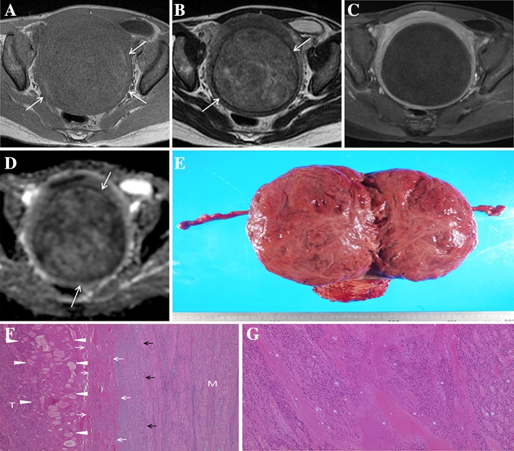
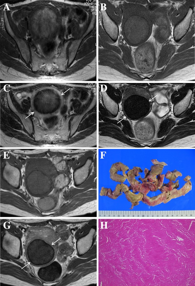
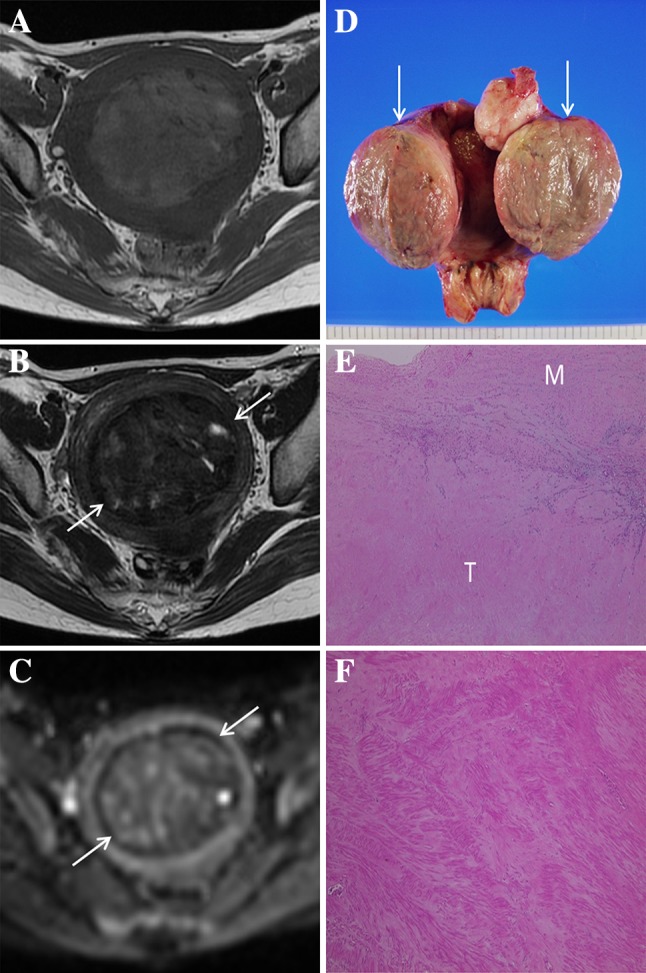
Venous infarction of a leiomyoma is known as red degeneration of leiomyoma (RDL) and can be a cause of acute abdomen. Although magnetic resonance imaging (MRI) is the only modality that can depict the inner condition of a leiomyoma, the typical MR findings of RDL are sometimes identified incidentally even in asymptomatic patients. The purpose of this study is to clarify common pathological findings of uterine tumors preoperatively diagnosed as RDL by MRI.

METHODS

We diagnosed 28 cases of RDL by MRI from March 2007 to April 2015. The ten lesions subjected to pathological analysis after resection were included in the study and reviewed by a gynecological pathologist. The average time from MRI to operation was 4.7 months.

RESULTS

The typical beefy-red color was not observed on the cut surface of the tumor except in one tumor resected during the acute phase. All lesions diagnosed as RDL by MRI had common pathological findings consistent with red degeneration of leiomyoma, including coagulative necrosis. Other common pathological features of RDL besides extensive coagulative necrosis appear to be a lack of inflammatory cell infiltrate or hemorrhage in the entire lesion.

CONCLUSIONS

Although RDL is known to cause acute abdomen, its typical MR findings can be observed even in asymptomatic patients in a condition that manifests long after red degeneration. The characteristic pathological findings in both the acute phase and the chronic phase that we found in this study, along with radiology reports, will be helpful references for gynecologists and pathologists in suspecting a history of red degeneration and confirming the diagnosis.

摘要

目的

子宫肌瘤静脉梗死又称子宫肌瘤红色变性(RDL),可引起急性腹痛。虽然磁共振成像(MRI)是唯一能显示子宫肌瘤内部情况的方法,但即使在无症状患者中,也会偶然发现 RDL 的典型 MRI 表现。本研究旨在阐明术前通过 MRI 诊断为 RDL 的子宫肌瘤的常见病理表现。

方法

我们从 2007 年 3 月至 2015 年 4 月通过 MRI 诊断了 28 例 RDL。纳入了 10 例经病理分析后切除的病变,并由妇科病理学家进行了回顾性分析。MRI 到手术的平均时间为 4.7 个月。

结果

除了在急性期切除的一个肿瘤外,肿瘤的切面未见典型的牛肉红色。所有通过 MRI 诊断为 RDL 的病变均具有与子宫肌瘤红色变性一致的常见病理表现,包括凝固性坏死。除广泛凝固性坏死外,RDL 的其他常见病理特征似乎是整个病变缺乏炎症细胞浸润或出血。

结论

虽然 RDL 可引起急性腹痛,但即使在红色变性后很长时间才出现症状的无症状患者中,也可观察到其典型的 MRI 表现。我们在本研究中发现的急性期和慢性期的特征性病理表现,以及放射学报告,将有助于妇科医生和病理学家怀疑红色变性病史并确认诊断。

https://cdn.ncbi.nlm.nih.gov/pmc/blobs/5c79/5486831/81d8eea3fe07/261_2017_1126_Fig3_HTML.jpg
https://cdn.ncbi.nlm.nih.gov/pmc/blobs/5c79/5486831/a84b5a7e6fb1/261_2017_1126_Fig1_HTML.jpg
https://cdn.ncbi.nlm.nih.gov/pmc/blobs/5c79/5486831/e029d7b0d42e/261_2017_1126_Fig2_HTML.jpg
https://cdn.ncbi.nlm.nih.gov/pmc/blobs/5c79/5486831/81d8eea3fe07/261_2017_1126_Fig3_HTML.jpg
https://cdn.ncbi.nlm.nih.gov/pmc/blobs/5c79/5486831/a84b5a7e6fb1/261_2017_1126_Fig1_HTML.jpg
https://cdn.ncbi.nlm.nih.gov/pmc/blobs/5c79/5486831/e029d7b0d42e/261_2017_1126_Fig2_HTML.jpg
https://cdn.ncbi.nlm.nih.gov/pmc/blobs/5c79/5486831/81d8eea3fe07/261_2017_1126_Fig3_HTML.jpg

相似文献

1
Pathological findings of uterine tumors preoperatively diagnosed as red degeneration of leiomyoma by MRI.术前 MRI 诊断为子宫肌瘤红色变性的子宫肿瘤的病理发现。
Abdom Radiol (NY). 2017 Jul;42(7):1825-1831. doi: 10.1007/s00261-017-1126-3.
2
Evaluation of Red Degeneration of Uterine Leiomyoma with Susceptibility-weighted MR Imaging.磁共振磁敏感加权成像评价子宫肌瘤红色变性。
Magn Reson Med Sci. 2019 Apr 10;18(2):158-162. doi: 10.2463/mrms.mp.2018-0074. Epub 2018 Oct 1.
3
Red degeneration of uterine leiomyoma: MR appearance.子宫平滑肌瘤红色变性:磁共振成像表现
J Comput Assist Tomogr. 1994 Nov-Dec;18(6):925-8. doi: 10.1097/00004728-199411000-00014.
4
MR imaging of atypical fibroids.磁共振成像在非典型纤维瘤中的应用。
Abdom Radiol (NY). 2016 Dec;41(12):2332-2349. doi: 10.1007/s00261-016-0935-0.
5
A subserosal, pedunculated, multilocular uterine leiomyoma with ovarian tumor-like morphology and histological architecture of adenomatoid tumors: a case report and review of the literature.一例具有腺瘤样瘤的卵巢肿瘤样形态和组织学结构的浆膜下带蒂多房性子宫平滑肌瘤:病例报告及文献复习
J Med Case Rep. 2016 Dec 20;10(1):352. doi: 10.1186/s13256-016-1167-1.
6
Diagnostic Accuracy of Ultrasound, Contrast-enhanced CT, and Conventional MRI for Differentiating Leiomyoma From Leiomyosarcoma.超声、对比增强CT及传统MRI鉴别平滑肌瘤与平滑肌肉瘤的诊断准确性
Acad Radiol. 2016 Oct;23(10):1290-7. doi: 10.1016/j.acra.2016.06.004. Epub 2016 Jul 7.
7
MR imaging findings of unusual leiomyoma and malignant uterine myometrial tumors: what the radiologist should know.磁共振成像对罕见的平滑肌瘤和恶性子宫平滑肌肿瘤的表现:放射科医生应该知道的。
Jpn J Radiol. 2021 Jun;39(6):527-539. doi: 10.1007/s11604-021-01096-7. Epub 2021 Jan 31.
8
Utility of Clinical Parameters and Multiparametric MRI as Predictive Factors for Differentiating Uterine Sarcoma From Atypical Leiomyoma.临床参数和多参数 MRI 在鉴别子宫肉瘤与非典型平滑肌瘤中的预测价值。
Acad Radiol. 2018 Aug;25(8):993-1002. doi: 10.1016/j.acra.2018.01.002. Epub 2018 Feb 13.
9
Unusual appearances of uterine leiomyomas: MR imaging findings and their histopathologic backgrounds.子宫平滑肌瘤的不寻常表现:磁共振成像结果及其组织病理学背景
Radiographics. 1999 Oct;19 Spec No:S131-45. doi: 10.1148/radiographics.19.suppl_1.g99oc04s131.
10
[Uterine leiomyoma embolization: Role of power Doppler ultrasonography].[子宫平滑肌瘤栓塞术:能量多普勒超声检查的作用]
Tani Girisim Radyol. 2003 Jun;9(2):240-5.

引用本文的文献

1
Multiparametric MRI manifestations of the spontaneous intratumoral coagulative necrosis in HCC.肝细胞癌内自发肿瘤性凝固性坏死的多参数 MRI 表现。
Abdom Radiol (NY). 2024 Jul;49(7):2198-2208. doi: 10.1007/s00261-024-04355-z. Epub 2024 May 17.
2
Acute abdomen by red degeneration of a parasitic leiomyoma: A case report and literature review.寄生性平滑肌瘤红色变性致急腹症:一例报告及文献复习
Radiol Case Rep. 2024 Jan 27;19(4):1533-1536. doi: 10.1016/j.radcr.2024.01.031. eCollection 2024 Apr.
3
Uterine myxoid leiomyosarcoma initially showing low signal intensity on T2 weighted images: A case report.

本文引用的文献

1
Pathology and pathophysiology of uterine smooth-muscle tumors.子宫平滑肌肿瘤的病理学与病理生理学
Environ Health Perspect. 2000 Oct;108 Suppl 5:779-84. doi: 10.1289/ehp.00108s5779.
2
Unusual appearances of uterine leiomyomas: MR imaging findings and their histopathologic backgrounds.子宫平滑肌瘤的不寻常表现:磁共振成像结果及其组织病理学背景
Radiographics. 1999 Oct;19 Spec No:S131-45. doi: 10.1148/radiographics.19.suppl_1.g99oc04s131.
3
Uterine leiomyomas: histopathologic features, MR imaging findings, differential diagnosis, and treatment.
子宫黏液性平滑肌肉瘤在T2加权图像上最初表现为低信号强度:一例报告。
Radiol Case Rep. 2023 Nov 29;19(2):700-705. doi: 10.1016/j.radcr.2023.11.034. eCollection 2024 Feb.
4
Imaging of Acute Abdominopelvic Pain in Pregnancy and Puerperium-Part II: Non-Obstetric Complications.妊娠和产褥期急性腹盆腔疼痛的影像学检查——第二部分:非产科并发症
Diagnostics (Basel). 2023 Sep 11;13(18):2909. doi: 10.3390/diagnostics13182909.
5
Uterine mesenchymal tumors: development and preliminary results of a magnetic resonance imaging (MRI) diagnostic algorithm.子宫间质肿瘤:磁共振成像(MRI)诊断算法的发展及初步结果。
Radiol Med. 2023 Jul;128(7):853-868. doi: 10.1007/s11547-023-01654-1. Epub 2023 Jun 14.
6
A case of primary aortic sarcoma with tumor infarction after stent graft placement.1例原发性主动脉肉瘤在置入支架型人工血管后出现肿瘤梗死。
Acta Radiol Open. 2021 Dec 7;10(11):20584601211063360. doi: 10.1177/20584601211063360. eCollection 2021 Nov.
7
Emergent transcatheter arterial embolization to control critical blood pressure fluctuation associated with hypercatecholaminemic crisis in a patient with an unruptured retroperitoneal paraganglioma.急诊经导管动脉栓塞术控制一名未破裂腹膜后副神经节瘤患者与高儿茶酚胺血症危象相关的严重血压波动。
Radiol Case Rep. 2021 Jun 8;16(8):2065-2071. doi: 10.1016/j.radcr.2021.05.018. eCollection 2021 Aug.
8
Uterine leiomyomas revisited with review of literature.子宫平滑肌瘤再探:文献复习。
Abdom Radiol (NY). 2021 Oct;46(10):4908-4926. doi: 10.1007/s00261-021-03126-4. Epub 2021 May 31.
9
Uterine fibroids: an update on current and emerging medical treatment options.子宫肌瘤:当前及新出现的医学治疗选择的最新进展
Ther Clin Risk Manag. 2019 Jan 23;15:157-178. doi: 10.2147/TCRM.S147318. eCollection 2019.
10
Evaluation of Red Degeneration of Uterine Leiomyoma with Susceptibility-weighted MR Imaging.磁共振磁敏感加权成像评价子宫肌瘤红色变性。
Magn Reson Med Sci. 2019 Apr 10;18(2):158-162. doi: 10.2463/mrms.mp.2018-0074. Epub 2018 Oct 1.
子宫平滑肌瘤:组织病理学特征、磁共振成像表现、鉴别诊断及治疗
Radiographics. 1999 Sep-Oct;19(5):1179-97. doi: 10.1148/radiographics.19.5.g99se131179.
4
Myomas and pregnancy.子宫肌瘤与妊娠
Obstet Gynecol Clin North Am. 1995 Dec;22(4):801-5.
5
MR appearance of hemorrhage in the brain.脑内出血的磁共振成像表现。
Radiology. 1993 Oct;189(1):15-26. doi: 10.1148/radiology.189.1.8372185.
6
Red degeneration of uterine leiomyoma: MR appearance.子宫平滑肌瘤红色变性:磁共振成像表现
J Comput Assist Tomogr. 1994 Nov-Dec;18(6):925-8. doi: 10.1097/00004728-199411000-00014.
7
Uterine leiomyoma. Incidence of degenerative change and a correlation of associated symptoms.子宫平滑肌瘤。退行性变的发生率及相关症状的相关性。
Obstet Gynecol. 1970 Mar;35(3):432-6.