He Xiao, Sun Dayu, Chen Siyu, Xu Haiwei
Southwest Hospital/Southwest Eye Hospital, Third Military Medical University, Chongqing, 400038, China.
Key Lab of Visual Damage and Regeneration & Restoration of Chongqing, Chongqing, 400038, China.
Oncotarget. 2017 May 9;8(19):32068-32082. doi: 10.18632/oncotarget.16643.
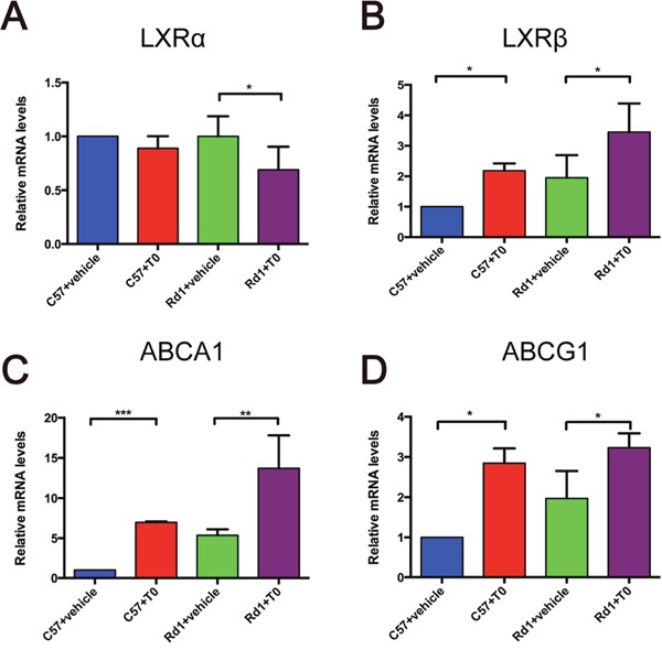
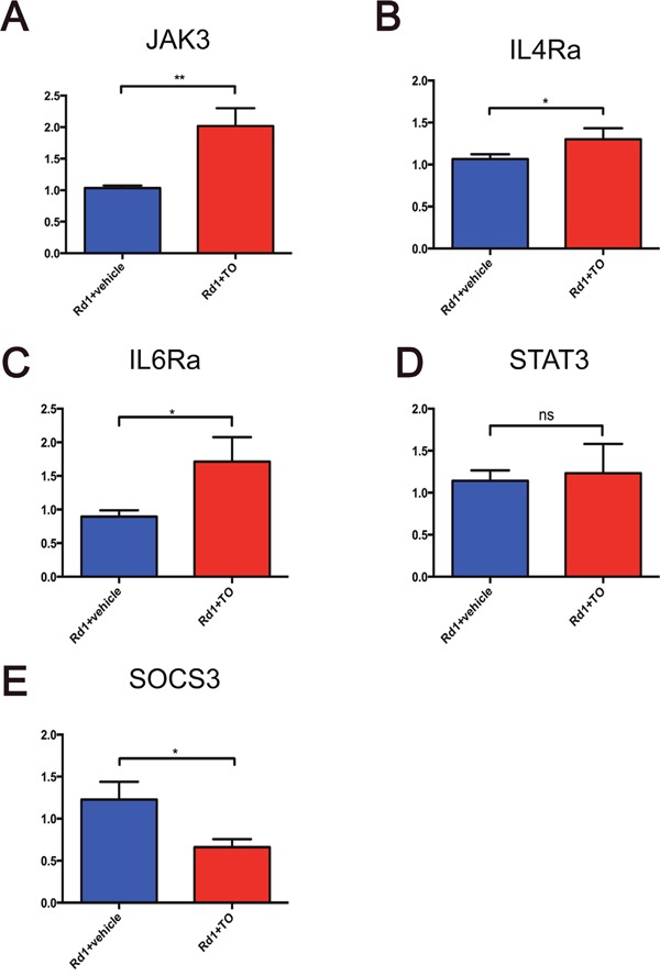
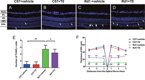
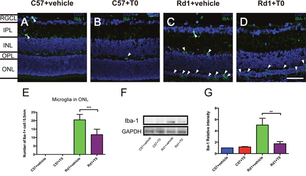
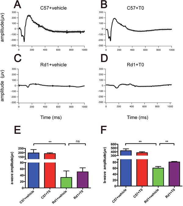
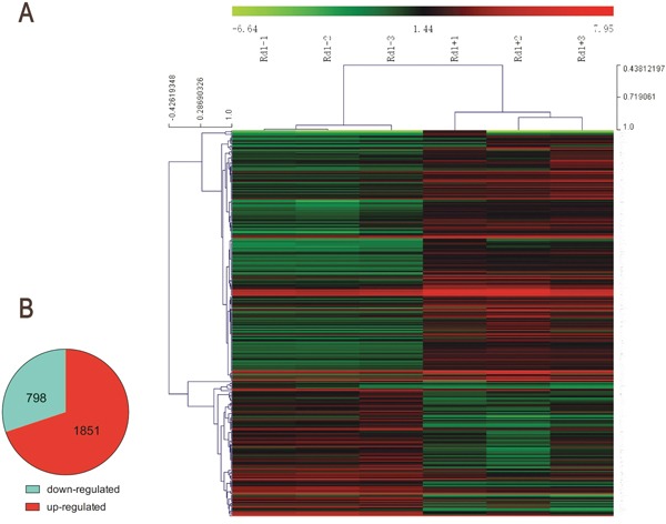
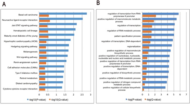
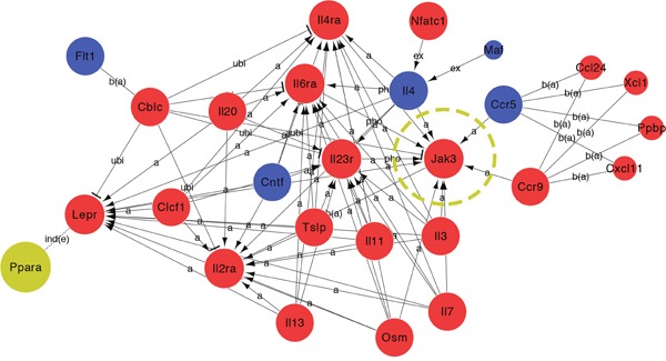
Retinal degeneration (RD), including retinitis pigmentosa (RP), is an inherited eye disease characterized by progressive degeneration of photoreceptors. Recently, immune cells, including microglia, Müller cells and astrocytes, in degenerative retina are demonstrated to play key roles in the development of RD and can be used as potential therapeutic targets. Liver X receptors (LXRs) are important immuno-inflammatory response transcription factors that have been reported to be a new potential therapeutic drug target for neurodegenerative diseases. However, the potential therapeutic utility of LXRs for RP has not been evaluated. In the present study, Pde6β (rd1) mice received intraperitoneal injections of T0901317 (T0, 50 mg/kg/d) or vehicle (2% DMSO) for 7 days with age-matched C57/BL6 mice as controls. The effect of T0 was examined by quantitating photoreceptor apoptosis, microglial density and the expression of inflammatory mediators; the underlying mechanisms were then explored with a microarray assay. T0 markedly delayed apoptosis of the photoreceptors, partially through suppressing the activation of microglia and the gliosis of Müller cells, and decreased the expression levels of IL-6, iNOS, COX-2 and ENG in rd1 mice; as a result, the visual function of T0-treated rd1 mice measured with electroretinograms (ERG) was preserved for a longer time than that of vehicle-treated rd1 mice. The microarray assay showed that the Janus kinase/Signal Transducer and Activator of Transcription (JAK-STAT) signaling pathway was significantly affected in the retina of rd1 mice with T0 treatment. Our data suggested that T0 modulated the immunologic function of glia cells in the degenerative retina through the JAK3/STAT pathway and delayed the apoptosis of photoreceptors.
视网膜变性(RD),包括色素性视网膜炎(RP),是一种遗传性眼病,其特征是光感受器进行性退化。最近,已证明退化视网膜中的免疫细胞,包括小胶质细胞、穆勒细胞和星形胶质细胞,在RD的发展中起关键作用,并且可作为潜在的治疗靶点。肝X受体(LXRs)是重要的免疫炎症反应转录因子,据报道是神经退行性疾病的一种新的潜在治疗药物靶点。然而,LXRs对RP的潜在治疗效用尚未得到评估。在本研究中,Pde6β(rd1)小鼠腹腔注射T0901317(T0,50mg/kg/d)或赋形剂(2%二甲亚砜),持续7天,以年龄匹配的C57/BL6小鼠作为对照。通过定量光感受器凋亡、小胶质细胞密度和炎症介质的表达来检测T0的作用;然后用微阵列分析探索其潜在机制。T0显著延迟了光感受器的凋亡,部分是通过抑制小胶质细胞的激活和穆勒细胞的胶质增生实现的,并降低了rd1小鼠中白细胞介素-6、诱导型一氧化氮合酶、环氧化酶-2和内皮糖蛋白的表达水平;结果,用视网膜电图(ERG)测量的T0处理的rd1小鼠的视觉功能比赋形剂处理的rd1小鼠的视觉功能保留的时间更长。微阵列分析表明,T0处理的rd1小鼠视网膜中Janus激酶/信号转导子和转录激活子(JAK-STAT)信号通路受到显著影响。我们的数据表明,T0通过JAK3/STAT途径调节退化视网膜中胶质细胞的免疫功能,并延迟光感受器的凋亡。