Townsend Michelle H, Anderson Michael D, Weagel Evita G, Velazquez Edwin J, Weber K Scott, Robison Richard A, O'Neill Kim L
Department of Microbiology and Molecular Biology, Brigham Young University, Provo, UT, USA.
Onco Targets Ther. 2017 Mar 30;10:1921-1932. doi: 10.2147/OTT.S128416. eCollection 2017.
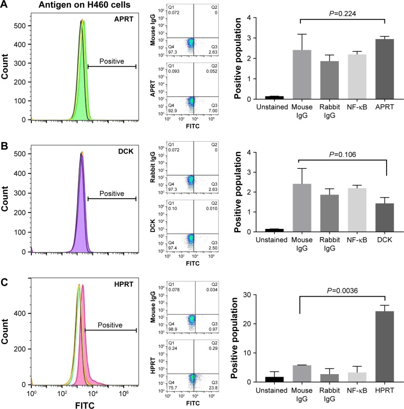
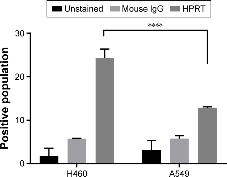
In both males and females, lung cancer is one of the most lethal cancers worldwide and accounts for >30% of cancer-related deaths. Despite advances in biomarker analysis and tumor characterization, there remains a need to find suitable biomarker antigen targets for treatment in late-stage lung cancer. Previous research on the salvage pathway enzyme TK1 shows a unique relationship with cancer patients as serum levels are raised according to cancer grade. To expand this analysis, the other salvage pathway enzymes were evaluated for possible upregulation within lung cancer. Adenine phosphoribosyltransferase, deoxycytidine kinase, and hypoxanthine guanine phosphoribosyltransferase (HPRT) were assessed for their presentation on two non-small-cell lung cancer cell lines NCI-H460 and A549. In the present study, we show that deoxycytidine kinase and adenine phosphoribosyltransferase have no significant relationship with the membrane of NCI-H460 cells. However, we found significant localization of HPRT to the membrane of NCI-H460 and A549 cells. When treated with anti-HPRT antibodies, the average fluorescence of the cell population increased by 24.3% and 12.9% in NCI-H460 and A549 cells, respectively, in comparison with controls. To ensure that expression was not attributed to cytoplasmic HPRT, confocal microscopy was performed to visualize HPRT binding on the plasma membrane. After staining NCI-H460 cells treated with both fluorescent antibodies and a membrane-specific dye, we observed direct overlap between HPRT and the membrane of the cancer cells. Additionally, gold-conjugated antibodies were used to label and quantify the amount of HPRT on the cell surface using scanning electron microscopy and energy-dispersive analysis X-ray. Further confirming HPRT presence, the gold weight percentage of the sample increased significantly when NCI-H460 cells were exposed to HPRT antibody (=0.012) in comparison with isotype controls. Our results show that HPRT is localized on the surface of these non-small-cell lung cancer cell lines.
在男性和女性中,肺癌都是全球最致命的癌症之一,占癌症相关死亡人数的30%以上。尽管生物标志物分析和肿瘤特征研究取得了进展,但仍需要寻找适合晚期肺癌治疗的生物标志物抗原靶点。先前对补救途径酶胸苷激酶1(TK1)的研究表明,它与癌症患者存在独特关系,因为血清水平会根据癌症分级升高。为了扩展这一分析,对其他补救途径酶在肺癌中可能的上调情况进行了评估。对腺嘌呤磷酸核糖转移酶、脱氧胞苷激酶和次黄嘌呤鸟嘌呤磷酸核糖转移酶(HPRT)在两种非小细胞肺癌细胞系NCI-H460和A549上的表达情况进行了评估。在本研究中,我们发现脱氧胞苷激酶和腺嘌呤磷酸核糖转移酶与NCI-H460细胞膜无显著关系。然而我们发现HPRT在NCI-H460和A549细胞膜上有显著定位。用抗HPRT抗体处理后,与对照组相比,NCI-H460和A549细胞群体的平均荧光分别增加了24.3%和12.9%。为确保表达并非源自细胞质HPRT,进行了共聚焦显微镜检查以观察HPRT在质膜上的结合情况。在用荧光抗体和膜特异性染料处理NCI-H460细胞后进行染色,我们观察到HPRT与癌细胞膜直接重叠。此外,使用金偶联抗体,通过扫描电子显微镜和能量色散X射线分析对细胞表面的HPRT量进行标记和定量。进一步证实了HPRT的存在,与同型对照相比,当NCI-H460细胞暴露于HPRT抗体时(P=0.012),样品的金重量百分比显著增加。我们的结果表明,HPRT定位于这些非小细胞肺癌细胞系的表面。