Attaye Ilias, Smulders Yvo M, de Waard Monique C, Oudemans-van Straaten Heleen M, Smit Bob, Van Wijhe Michiel H, Musters Rene J, Koolwijk Pieter, Spoelstra-de Man Angelique M E
Department of Intensive Care, VU University Medical Center, Amsterdam, The Netherlands.
Department of Physiology, VU University Medical Center, Amsterdam, The Netherlands.
Intensive Care Med Exp. 2017 Dec;5(1):22. doi: 10.1186/s40635-017-0135-4. Epub 2017 Apr 13.
Hyperoxia, an arterial oxygen pressure of more than 100 mmHg or 13% O, frequently occurs in hospitalized patients due to administration of supplemental oxygen. Increasing evidence suggests that hyperoxia induces vasoconstriction in the systemic (micro)circulation, potentially affecting organ perfusion. This study addresses effects of hyperoxia on viability, proliferative capacity, and on pathways affecting vascular tone in cultured human microvascular endothelial cells (hMVEC).
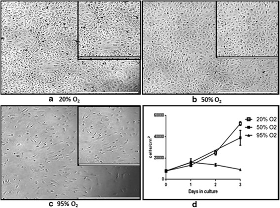
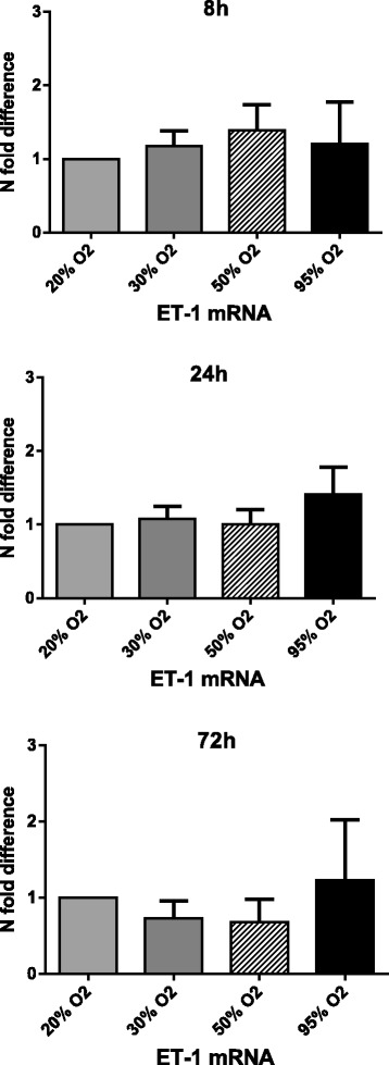
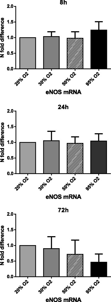
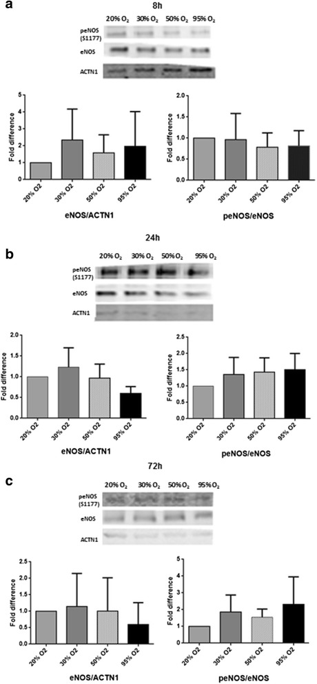
hMVEC of the systemic circulation were exposed to graded oxygen fractions of 20, 30, 50, and 95% O for 8, 24, and 72 h. These fractions correspond to 152, 228, 380, and 722 mmHg, respectively. Cell proliferation and viability was measured via a proliferation assay, peroxynitrite formation via anti-nitrotyrosine levels, endothelial nitric oxide synthase (eNOS), and endothelin-1 (ET-1) levels via q-PCR and western blot analysis.
Exposing hMVEC to 50 and 95% O for more than 24 h impaired cell viability and proliferation. Hyperoxia did not significantly affect nitrotyrosine levels, nor eNOS mRNA and protein levels, regardless of the exposure time or oxygen concentration used. Phosphorylation of eNOS at the serine 1177 (S1177) residue and ET-1 mRNA levels were also not significantly affected.
Exposure of isolated human microvascular endothelial cells to marked hyperoxia for more than 24 h decreases cell viability and proliferation. Our results do not support a role of eNOS mRNA and protein or ET-1 mRNA in the potential vasoconstrictive effects of hyperoxia on isolated hMVEC.
高氧,即动脉血氧分压超过100 mmHg或氧含量超过13%,在住院患者中因补充氧气的使用而经常发生。越来越多的证据表明,高氧会诱导全身(微)循环中的血管收缩,可能影响器官灌注。本研究探讨高氧对培养的人微血管内皮细胞(hMVEC)的活力、增殖能力以及影响血管张力的信号通路的作用。
将体循环的hMVEC暴露于20%、30%、50%和95%的梯度氧分压下8小时、24小时和72小时。这些氧分压分别对应152 mmHg、228 mmHg、380 mmHg和722 mmHg。通过增殖试验测量细胞增殖和活力,通过抗硝基酪氨酸水平测量过氧亚硝酸盐的形成,通过q-PCR和蛋白质印迹分析测量内皮型一氧化氮合酶(eNOS)和内皮素-1(ET-1)水平。
将hMVEC暴露于50%和95%的氧分压下超过24小时会损害细胞活力和增殖。无论暴露时间或氧浓度如何,高氧均未显著影响硝基酪氨酸水平、eNOS mRNA和蛋白质水平。eNOS丝氨酸1177(S1177)残基的磷酸化和ET-1 mRNA水平也未受到显著影响。
将分离的人微血管内皮细胞暴露于明显的高氧环境中超过24小时会降低细胞活力和增殖。我们的结果不支持eNOS mRNA和蛋白质或ET-1 mRNA在高氧对分离的hMVEC潜在血管收缩作用中的作用。