Department of Surgery, Comprehensive Cancer Center Vienna, Upper-GI-Service, GET-Unit, Medical University of Vienna, Vienna, Austria.
Department of Dermatology, University Hospital Zürich, University of Zurich, Zurich, Switzerland.
Ann Surg Oncol. 2017 Sep;24(9):2698-2706. doi: 10.1245/s10434-017-5858-7. Epub 2017 Apr 20.
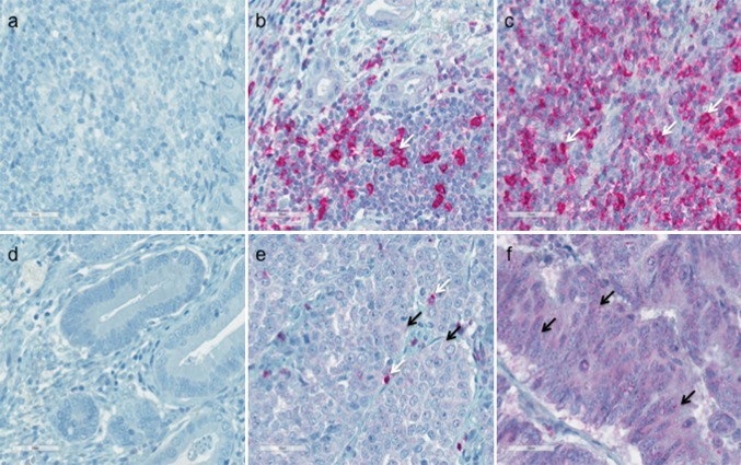
Despite recent advances in the therapy for adenocarcinoma of the esophagogastric junction (AEG), overall prognosis remains poor. Programmed cell death protein 1 (PD1) is a co-inhibitory receptor primarily expressed by T-cells. Tumor cells can escape anticancer immune responses by triggering the PD1 pathway. Moreover, PD1 receptor engagement on cancer cells may trigger tumor-intrinsic growth signals. This study aimed to evaluate the potential clinical relevance of PD1 expression by tumor-infiltrating lymphocytes (TILs) and cancer cells in the AEG.
Patients with AEG who underwent esophagectomy from 1992 to 2011 were included in the study. Expression of PD1was evaluated by immunohistochemistry and correlated with long-term overall survival (OS), disease-free survival (DFS), and various clinicopathologic parameters.
Tumor biospecimens from 168 patients were analyzed. In the analysis, 81% of the patients showed high tumoral frequencies (>5%) of PD1-expressing TILs (TIL-PD1), and 77% of patient tumors harbored high levels (>5%) of PD1 cancer cells (cancer-PD1). Expression of PD1 by TILs and cancer cells correlated significantly (p < 0.05) with patients' tumor stage and lymph node involvement. Compared with the patients who had low tumoral frequencies of PD1 TILs or cancer cells, the TIL-PD1 and cancer-PD1 patients demonstrated significantly reduced DFS in the univariate analysis (5-year DFS: 73.3 vs. 41.9%, log-rank 0.008 and 71.3 vs. 41.6%, p = 0.008, respectively). Additionally, the cancer-PD1 patients showed significantly decreased OS in the univariate analysis compared with the cancer-PD1 patients (5-year OS: 68.8 vs. 43.5%; p = 0.047). However, these correlations did not reach significance in the multivariate analysis.
The PD1 receptor is expressed by both TILs and cancer cells in AEG. High expression of PD1 is associated with advanced tumor stage and lymph node involvement, but not with survival.
尽管食管胃结合部腺癌(AEG)的治疗方法最近有所进展,但总体预后仍然较差。程序性细胞死亡蛋白 1(PD1)是一种主要在 T 细胞上表达的共抑制受体。肿瘤细胞可以通过触发 PD1 途径来逃避抗癌免疫反应。此外,癌细胞上 PD1 受体的结合可能会触发肿瘤内在的生长信号。本研究旨在评估肿瘤浸润淋巴细胞(TILs)和 AEG 中癌细胞 PD1 表达的潜在临床相关性。
纳入 1992 年至 2011 年间接受食管切除术的 AEG 患者。通过免疫组织化学评估 PD1 的表达,并将其与长期总生存率(OS)、无病生存率(DFS)和各种临床病理参数相关联。
分析了 168 名患者的肿瘤生物标本。在分析中,81%的患者显示 TIL-PD1(肿瘤中 PD1 表达 TIL 的高频率>5%),77%的患者肿瘤中存在高水平的 PD1 癌细胞(cancer-PD1)。TILs 和癌细胞中 PD1 的表达与患者的肿瘤分期和淋巴结受累显著相关(p<0.05)。与 TIL-PD1 和 cancer-PD1 患者肿瘤中 PD1 TILs 或癌细胞的低频率相比,TIL-PD1 和 cancer-PD1 患者在单因素分析中显示出显著降低的 DFS(5 年 DFS:73.3%比 41.9%,log-rank 0.008 和 71.3%比 41.6%,p=0.008)。此外,与 cancer-PD1 患者相比,cancer-PD1 患者在单因素分析中显示出显著降低的 OS(5 年 OS:68.8%比 43.5%;p=0.047)。然而,这些相关性在多因素分析中没有达到显著水平。
PD1 受体在 AEG 中的 TILs 和癌细胞中均有表达。PD1 的高表达与肿瘤分期和淋巴结受累有关,但与生存率无关。