Lim Bock, Yao Yu, Huang Alex Lin-I, Yap May Lin, Flierl Ulrike, Palasubramaniam Jathushan, Zaldivia Maria T K, Wang Xiaowei, Peter Karlheinz
Atherothrombosis & Vascular Biology, Baker IDI Heart and Diabetes Institute, Melbourne, Australia.
Department of Medicine Monash University, Melbourne, Australia.
Theranostics. 2017 Feb 26;7(5):1047-1061. doi: 10.7150/thno.18099. eCollection 2017.
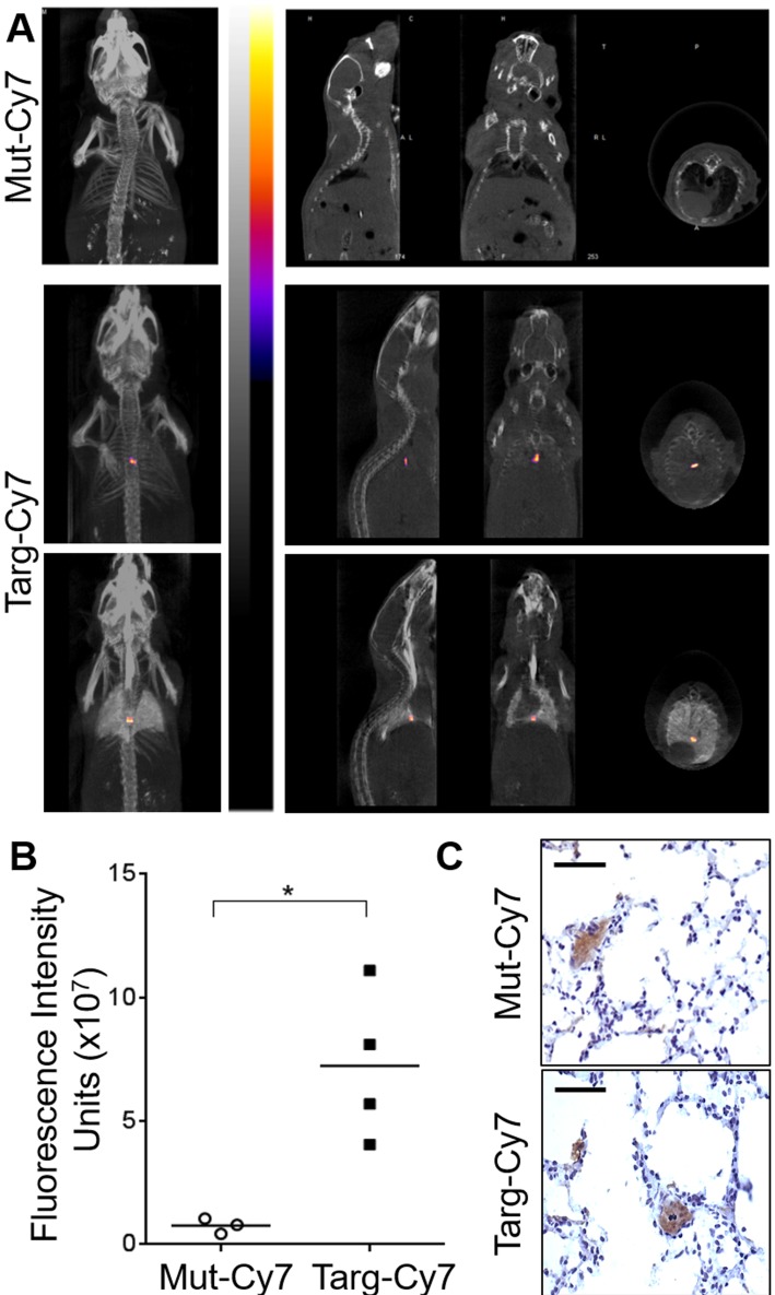
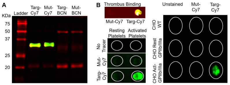
Progress in pharmaceutical development is highly-dependent on preclinical animal studies. Small animal imaging is invaluable for the identification of new disease markers and the evaluation of drug efficacy. Here, we report for the first time the use of a three-dimensional fluorescence bioimager called FLuorescence Emission Computed Tomography (FLECT) for the detection of a novel recombinant fluoroprobe that is safe, easily prepared on a large scale and stably stored prior to scan. This novel fluoroprobe (Targ-Cy7) comprises a single-chain antibody-fragment (scFv), which binds exclusively to activated-platelets, conjugated to a near-infrared (NIR) dye, Cy7, for detection. Upon mouse carotid artery injury, the injected fluoroprobe circulates and binds within the platelet-rich thrombus. This specific binding of the fluoroprobe to the thrombus, compared to its non-targeting control-fluoroprobe, is detected by the FLECT imager. The analyzed FLECT image quantifies the NIR signal and localizes it to the site of vascular injury. The detected fluorescence is further verified using a two-dimensional IVIS Lumina scanner, where significant NIR fluorescence is detected at the thrombotic site, and , at the injured carotid artery. Furthermore, fluorescence levels in various organs have also been quantified for biodistribution, with the highest fluoroprobe uptake shown to be in the injured artery. Subsequently, this live animal imaging technique is successfully employed to monitor the response of the induced thrombus to treatment over time. This demonstrates the potential of using longitudinal FLECT scanning to examine the efficacy of candidate drugs in preclinical settings. Besides intravascular thrombosis, we have shown that this non-invasive FLECT-imaging can also detect pulmonary embolism. Overall, this report describes a novel fluorescence-based preclinical imaging modality that uses an easy-to-prepare and non-radioactive recombinant fluoroprobe. This represents a unique tool to study mechanisms of thromboembolic diseases and it will strongly facilitate the testing of antithrombotic drugs. Furthermore, the non-radiation nature, low-cost, high sensitivity, and the rapid advancement of optical scanning technologies make this fluorescence imaging an attractive development for future clinical applications.
药物研发的进展高度依赖临床前动物研究。小动物成像对于识别新的疾病标志物和评估药物疗效具有重要价值。在此,我们首次报告使用一种名为荧光发射计算机断层扫描(FLECT)的三维荧光生物成像仪来检测一种新型重组荧光探针,该探针安全、易于大规模制备且在扫描前可稳定保存。这种新型荧光探针(Targ-Cy7)由一个单链抗体片段(scFv)组成,该片段专门与活化血小板结合,并与一种近红外(NIR)染料Cy7偶联用于检测。在小鼠颈动脉损伤后,注入的荧光探针在富含血小板的血栓内循环并结合。与非靶向对照荧光探针相比,荧光探针与血栓的这种特异性结合可通过FLECT成像仪检测到。分析的FLECT图像对近红外信号进行定量,并将其定位到血管损伤部位。使用二维IVIS Lumina扫描仪进一步验证检测到的荧光,在血栓形成部位以及受伤的颈动脉处检测到显著的近红外荧光。此外,还对各个器官中的荧光水平进行了定量以研究生物分布,显示荧光探针在受伤动脉中的摄取量最高。随后,这种活体动物成像技术成功用于监测诱导血栓随时间对治疗的反应。这证明了使用纵向FLECT扫描在临床前环境中检查候选药物疗效的潜力。除了血管内血栓形成外,我们还表明这种非侵入性FLECT成像还可以检测肺栓塞。总体而言,本报告描述了一种基于新型荧光的临床前成像模式,该模式使用易于制备且无放射性的重组荧光探针。这代表了一种研究血栓栓塞性疾病机制的独特工具,将极大地促进抗血栓药物的测试。此外,非辐射性质、低成本、高灵敏度以及光学扫描技术的快速发展使这种荧光成像成为未来临床应用的一个有吸引力的发展方向。