Choubey Lisha, Collette Jantzen C, Smith Karen Müller
Department of Biology, University of Louisiana at Lafayette, United States of America.
PeerJ. 2017 Apr 18;5:e3173. doi: 10.7717/peerj.3173. eCollection 2017.
Fibroblast growth factors (FGFs) and their receptors (FGFRs) have numerous functions in the developing and adult central nervous system (CNS). For example, the FGFR1 receptor is important for proliferation and fate specification of radial glial cells in the cortex and hippocampus, oligodendrocyte proliferation and regeneration, midline glia morphology and soma translocation, Bergmann glia morphology, and cerebellar morphogenesis. In addition, FGFR1 signaling in astrocytes is required for postnatal maturation of interneurons expressing parvalbumin (PV). FGFR1 is implicated in synapse formation in the hippocampus, and alterations in the expression of and its ligand, accompany major depression. Understanding which cell types express during development may elucidate its roles in normal development of the brain as well as illuminate possible causes of certain neuropsychiatric disorders.
Here, we used a BAC transgenic reporter line to trace expression in the developing postnatal murine CNS. The specific transgenic line employed was created by the GENSAT project, , and includes a gene encoding enhanced green fluorescent protein () under the regulation of the promoter, to trace expression in the developing CNS. Unbiased stereological counts were performed for several cell types in the cortex and hippocampus.
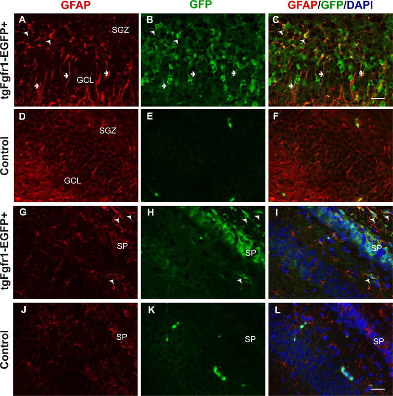
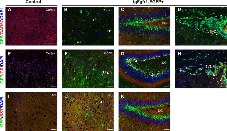
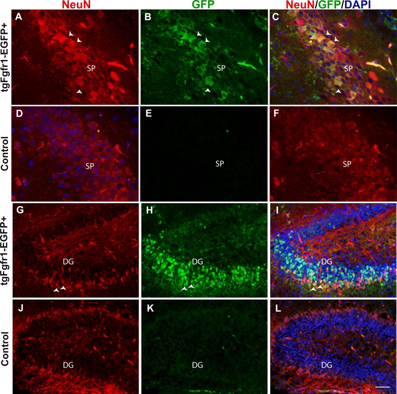
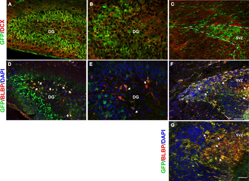
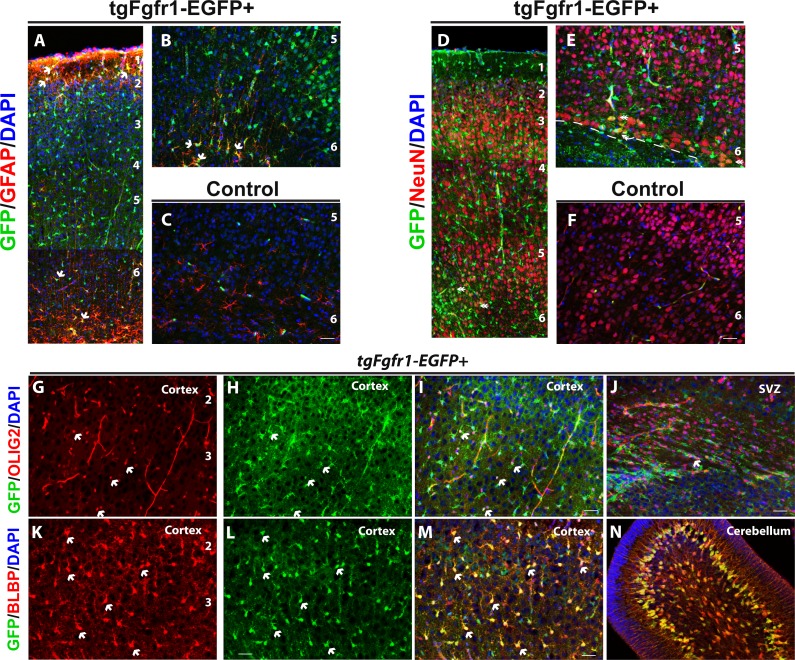
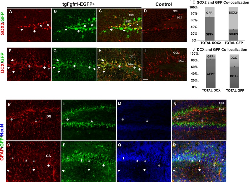
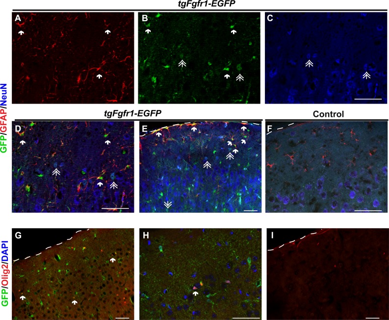
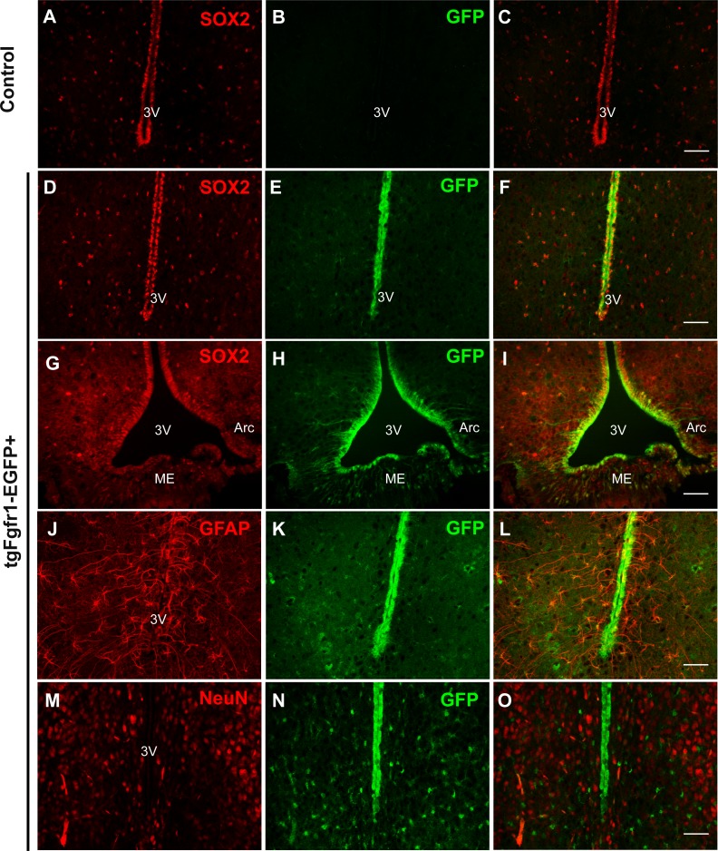
This model reveals that is primarily expressed in glial cells, in both astrocytes and oligodendrocytes, along with some neurons. Dual labeling experiments indicate that the proportion of GFP+ (+) cells that are also GFAP+ increases from postnatal day 7 (P7) to 1 month, illuminating dynamic changes in expression during postnatal development of the cortex. In postnatal neurogenic areas, GFP expression was also observed in SOX2, doublecortin (DCX), and brain lipid-binding protein (BLBP) expressing cells. is also highly expressed in DCX positive cells of the dentate gyrus (DG), but not in the rostral migratory stream. driven GFP was also observed in tanycytes and GFAP+ cells of the hypothalamus, as well as in Bergmann glia and astrocytes of the cerebellum.
The mouse model expresses GFP that is congruent with known functions of FGFR1, including hippocampal development, glial cell development, and stem cell proliferation. Understanding which cell types express may elucidate its role in neuropsychiatric disorders and brain development.
成纤维细胞生长因子(FGFs)及其受体(FGFRs)在发育中的和成年的中枢神经系统(CNS)中具有多种功能。例如,FGFR1受体对于皮质和海马中放射状胶质细胞的增殖和命运决定、少突胶质细胞的增殖和再生、中线胶质细胞形态和胞体移位、伯格曼胶质细胞形态以及小脑形态发生都很重要。此外,表达小白蛋白(PV)的中间神经元的出生后成熟需要星形胶质细胞中的FGFR1信号传导。FGFR1与海马中的突触形成有关,其表达及其配体的改变与重度抑郁症相伴。了解在发育过程中哪些细胞类型表达FGFR1可能有助于阐明其在大脑正常发育中的作用,并揭示某些神经精神疾病的可能病因。
在这里,我们使用了一种BAC转基因报告系来追踪出生后发育中的小鼠中枢神经系统中FGFR1的表达。所使用的特定转基因系是由GENSAT项目创建的,即BAC-FGFR1-EGFP,并且包括一个在FGFR1启动子调控下编码增强型绿色荧光蛋白(EGFP)的基因,以追踪发育中的中枢神经系统中FGFR1的表达。对皮质和海马中的几种细胞类型进行了无偏倚的立体学计数。
该模型显示,FGFR1主要在胶质细胞中表达,包括星形胶质细胞和少突胶质细胞,以及一些神经元。双重标记实验表明,也是GFAP+的GFP+(EGFP+)细胞的比例从出生后第7天(P7)到1个月增加,这揭示了皮质出生后发育过程中FGFR1表达的动态变化。在出生后神经发生区域,在表达SOX2、双皮质素(DCX)和脑脂质结合蛋白(BLBP)的细胞中也观察到了GFP表达。FGFR1在齿状回(DG)的DCX阳性细胞中也高度表达,但在吻侧迁移流中不表达。在室管膜细胞和下丘脑的GFAP+细胞中,以及在小脑的伯格曼胶质细胞和星形胶质细胞中也观察到了FGFR1驱动的GFP。
FGFR1小鼠模型表达的GFP与FGFR1的已知功能一致,包括海马发育、胶质细胞发育和干细胞增殖。了解哪些细胞类型表达FGFR1可能有助于阐明其在神经精神疾病和大脑发育中的作用。