Cerqueira Emanuella Rd, Valente Neusa, Sotto Mírian Nacagami, Romiti Ricardo
Department of Dermatology, University of São Paulo Medical School, São Paulo, Brazil.
Department of Pathology, University of São Paulo Medical School, São Paulo, Brazil.
Int J Trichology. 2016 Oct-Dec;8(4):197-202. doi: 10.4103/0974-7753.203179.
Frontal fibrosing alopecia (FFA) is a disorder characterized by progressive cicatricial alopecia (CA). Its classification as a clinical variant of lichen planopilaris (LPP) or as a unique disorder is controversial. The presence of Langerhans cells within the bulge area and the sebaceous epithelium and the presence of lymphocytic infiltrate in this area in CA have led to a series of hypotheses, although limited, about their development. To our knowledge, scarce is the literature demonstrating immunoanalytical studies comparing both disorders.
The authors sought to describe diagnostic findings, comorbidities, and immunopathological features of female patients with FFA as compared to LPP.
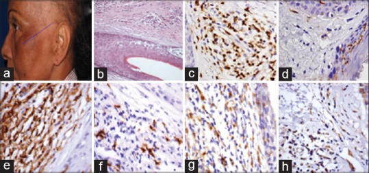
This retrospective single-center study included patients given the diagnosis of FFA or LPP. The LPP activity index was used to evaluate objective signs and subjective symptoms. Biopsy specimens were obtained from active, inflammatory areas of the scalp, and the inflammatory infiltrate intensity and quality were compared. Direct immunofluorescence for IgA, IgM, and IgG and immunohistochemistry to demonstrate the expression of CD1a, CD3, CD4, CD8, CD68, and 2,3-dioxygenase indoleamine were performed.
Twenty female patients (10 patients with FFA and 10 patients with LPP) were included in the study. Histopathological findings evidenced reduced number of hair follicles and perifollicular fibrosis in both disorders. Immunofluorescence findings resulted positive in 50% of FFA cases and 40% of LPP cases.
Although clinically different, our findings suggest that there are, to date, no histological or immunological findings that allow us to accurately separate these two forms of scarring alopecia, namely FFA and LPP.
额部纤维性秃发(FFA)是一种以进行性瘢痕性秃发(CA)为特征的疾病。它被归类为扁平苔藓样秃发(LPP)的临床变体还是一种独特的疾病存在争议。CA患者毛囊隆突区和皮脂腺上皮内朗格汉斯细胞的存在以及该区域淋巴细胞浸润的存在引发了一系列关于其发病机制的假说,尽管这些假说有限。据我们所知,很少有文献展示比较这两种疾病的免疫分析研究。
作者试图描述FFA女性患者与LPP患者相比的诊断结果、合并症和免疫病理特征。
这项回顾性单中心研究纳入了被诊断为FFA或LPP的患者。使用LPP活动指数评估客观体征和主观症状。从头皮的活跃炎症区域获取活检标本,并比较炎症浸润的强度和性质。进行了针对IgA、IgM和IgG的直接免疫荧光以及用于证明CD1a、CD3、CD4、CD8、CD68和吲哚胺2,3 -双加氧酶表达的免疫组织化学检测。
该研究纳入了20名女性患者(10名FFA患者和10名LPP患者)。组织病理学结果表明两种疾病中毛囊数量均减少且毛囊周围纤维化。免疫荧光结果在50%的FFA病例和40%的LPP病例中呈阳性。
尽管在临床上有所不同,但我们的研究结果表明,迄今为止,尚无组织学或免疫学发现能够使我们准确区分这两种瘢痕性秃发形式,即FFA和LPP。