Hellstrøm Torgeir, Kaufmann Tobias, Andelic Nada, Soberg Helene L, Sigurdardottir Solrun, Helseth Eirik, Andreassen Ole A, Westlye Lars T
Department of Physical Medicine and Rehabilitation, Oslo University Hospital, Oslo, Norway.
Faculty of Medicine, Institute of Clinical Medicine, University of Oslo, Oslo, Norway.
Front Neurol. 2017 Apr 10;8:125. doi: 10.3389/fneur.2017.00125. eCollection 2017.
Accurate outcome prediction models for patients with mild traumatic brain injury (MTBI) are key for prognostic assessment and clinical decision-making. Using multivariate machine learning, we tested the unique and added predictive value of (1) magnetic resonance imaging (MRI)-based brain morphometric and volumetric characterization at 4-week postinjury and (2) demographic, preinjury, injury-related, and postinjury variables on 12-month outcomes, including global functioning level, postconcussion symptoms, and mental health in patients with MTBI.
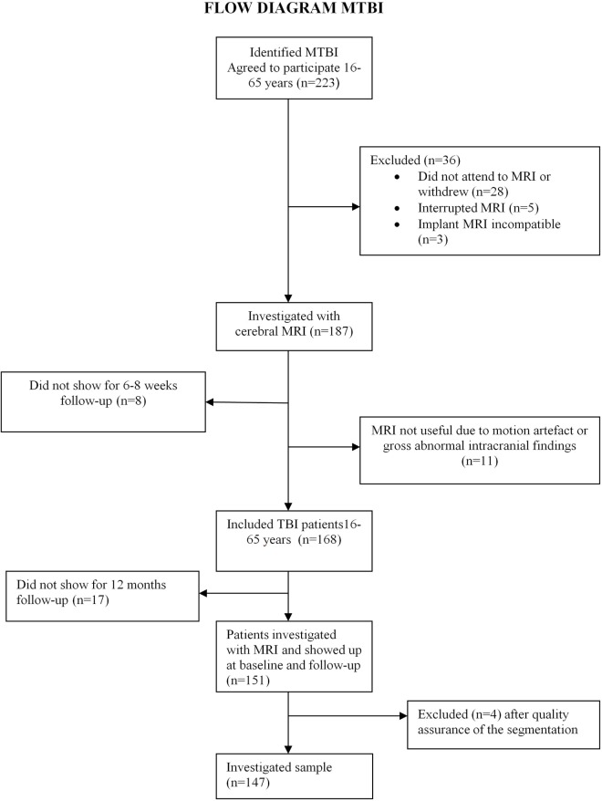
A prospective, cohort study of patients ( = 147) aged 16-65 years with a 12-month follow-up. T1-weighted 3 T MRI data were processed in FreeSurfer, yielding accurate cortical reconstructions for surface-based analyses of cortical thickness, area, and volume, and brain segmentation for subcortical and global brain volumes. The 12-month outcome was defined as a composite score using a principal component analysis including the Glasgow Outcome Scale Extended, Rivermead Postconcussion Questionnaire, and Patient Health Questionnaire-9. Using leave-one-out cross-validation and permutation testing, we tested and compared three prediction models: (1) MRI model, (2) clinical model, and (3) MRI and clinical combined.
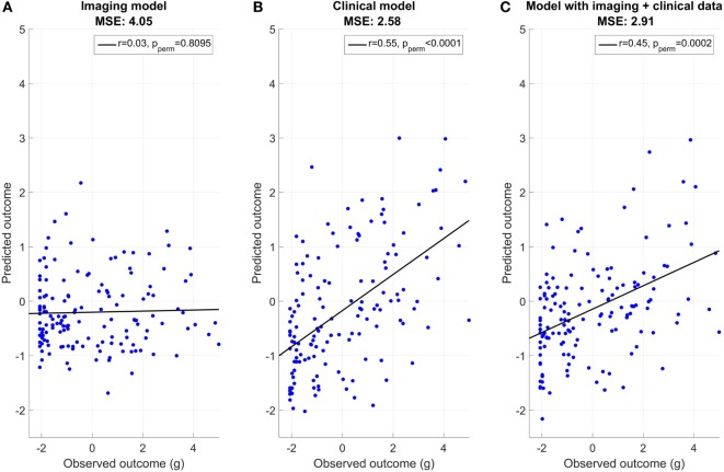
We found a strong correlation between observed and predicted outcomes for the clinical model ( = 0.55, < 0.001). The MRI model performed at the chance level ( = 0.03, = 0.80) and the combined model ( = 0.45, < 0.002) were slightly weaker than the clinical model. Univariate correlation analyses revealed the strongest association with outcome for postinjury factors of posttraumatic stress (Posttraumatic Symptom Scale-10, = 0.61), psychological distress (Hospital Anxiety and Depression Scale, = 0.52), and widespread pain ( = 0.43) assessed at 8 weeks.
We found no added predictive value of MRI-based measures of brain cortical morphometry and subcortical volumes over and above demographic and clinical features.
准确的轻度创伤性脑损伤(MTBI)患者预后预测模型是预后评估和临床决策的关键。我们使用多变量机器学习方法,测试了(1)伤后4周基于磁共振成像(MRI)的脑形态计量学和体积特征,以及(2)人口统计学、伤前、损伤相关和伤后变量对MTBI患者12个月预后的独特和附加预测价值,这些预后包括整体功能水平、脑震荡后症状和心理健康。
对147例年龄在16 - 65岁的患者进行前瞻性队列研究,并进行12个月的随访。T1加权3T MRI数据在FreeSurfer中进行处理,生成准确的皮质重建图像,用于基于表面的皮质厚度、面积和体积分析,以及用于皮质下和全脑体积的脑分割。12个月的预后通过主成分分析定义为一个综合评分,包括格拉斯哥扩展预后量表、Rivermead脑震荡后问卷和患者健康问卷-9。使用留一法交叉验证和置换检验,我们测试并比较了三个预测模型:(1)MRI模型,(2)临床模型,以及(3)MRI与临床联合模型。
我们发现临床模型的观察结果与预测结果之间存在强相关性(r = 0.55,p < 0.001)。MRI模型的表现处于随机水平(r = 0.03,p = 0.80),联合模型(r = 0.45,p < 0.002)略弱于临床模型。单变量相关性分析显示,伤后8周评估的创伤后应激(创伤后症状量表-10,r = 0.61)、心理困扰(医院焦虑抑郁量表,r = 0.52)和广泛疼痛(r = 0.43)等因素与预后的关联最强。
我们发现,除了人口统计学和临床特征外,基于MRI的脑皮质形态计量学和皮质下体积测量没有附加的预测价值。