Department of Radiology, Hacettepe University School of Medicine, Ankara, Turkey.
Balkan Med J. 2017 Aug 4;34(4):371-373. doi: 10.4274/balkanmedj.2016.0292. Epub 2017 Apr 13.
Cerebral venous air embolism is a severe clinical condition related to an unfavourable outcome in patients with neurological impairment. Cerebral venous air embolism may occur secondarily to arterial or venous interventions. A rare mechanism of cerebral venous air embolism is retrograde embolism, which is characterized by gas flow in a direction that is opposite to that of the normal blood flow.
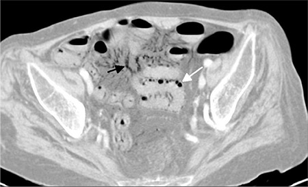
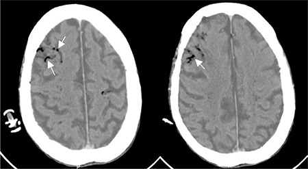
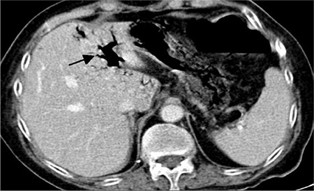
A 69-year-old female was admitted to our hospital with shortness of breath and abdominal pain. Abdominal computed tomography revealed intramural gas in the bowel and free gas in the mesenteric veins and portal vein. Cranial computed tomography, which was performed due to impaired consciousness, demonstrated cerebral air embolism with the appearance of a gyriform pattern. A bedside echocardiography and chest computed tomography revealed no evidence of right-to-left shunt.
Cerebral venous air embolism may occur after pneumatosis intestinalis by a retrograde flow of air from the mesenteric veins and portal vein. Low cardiac output and supine position are contributing factors for a retrograde flow of air bubbles into the venous circulation of the brain.
脑静脉气栓是一种严重的临床情况,与神经功能障碍患者的不良预后相关。脑静脉气栓可继发于动脉或静脉介入。脑静脉气栓的一种罕见机制是逆行性栓塞,其特征为气体流向与正常血流方向相反。
一名 69 岁女性因呼吸急促和腹痛入院。腹部 CT 显示肠壁内气体和肠系膜静脉及门静脉内游离气体。因意识障碍进行的颅脑 CT 显示脑气栓,呈脑回样模式。床边超声心动图和胸部 CT 未发现右向左分流证据。
肠气肿可通过空气从肠系膜静脉和门静脉逆行流动导致脑静脉气栓。低心输出量和仰卧位是空气气泡逆行流入脑部静脉循环的促成因素。