• 文献检索
  • 文档翻译
  • 深度研究
  • 学术资讯
  • Suppr Zotero 插件Zotero 插件
  • 邀请有礼
  • 套餐&价格
  • 历史记录
应用&插件
Suppr Zotero 插件Zotero 插件浏览器插件Mac 客户端Windows 客户端微信小程序
定价
高级版会员购买积分包购买API积分包
服务
文献检索文档翻译深度研究API 文档MCP 服务
关于我们
关于 Suppr公司介绍联系我们用户协议隐私条款
关注我们

Suppr 超能文献

核心技术专利:CN118964589B侵权必究
粤ICP备2023148730 号-1Suppr @ 2026

文献检索

告别复杂PubMed语法,用中文像聊天一样搜索,搜遍4000万医学文献。AI智能推荐,让科研检索更轻松。

立即免费搜索

文件翻译

保留排版,准确专业,支持PDF/Word/PPT等文件格式,支持 12+语言互译。

免费翻译文档

深度研究

AI帮你快速写综述,25分钟生成高质量综述,智能提取关键信息,辅助科研写作。

立即免费体验

内镜超声检查预测肝硬化患者早期食管静脉曲张出血:一例报告。

Endoscopic ultrasonography predicts early esophageal variceal bleeding in liver cirrhosis: A case report.

作者信息

Men Changjun, Zhang Guoliang

机构信息

Digestive Department, Tianjin First Center Hospital, Tianjin, P.R. China.

出版信息

Medicine (Baltimore). 2017 Apr;96(17):e6749. doi: 10.1097/MD.0000000000006749.

DOI:10.1097/MD.0000000000006749
PMID:28445299
原文链接:https://pmc.ncbi.nlm.nih.gov/articles/PMC5413264/
Abstract

RATIONALE

Bleeding esophageal and gastric varices constitute a serious complication in liver cirrhosis. Previous studies have shown that endoscopic ultrasonography (EUS) can be used to predict early esophageal variceal bleeding in liver cirrhosis.

PATIENT CONCERNS

We report a case of a 46-year-old man with hepatitis B liver cirrhosis (CTP score, 5; Child-Pugh class, A) who was admitted to our hospital due to a decreased appetite lasting 1 week.

DIAGNOSIS

He was initially diagnosed with decompensated hepatitis B cirrhosis; an abdominal computed tomography (CT) scan indicated a diagnosis of liver cirrhosis and portal hypertension (PHT).

INTERVENTIONS

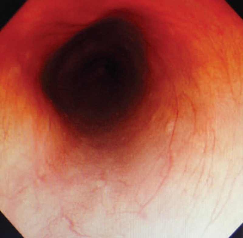
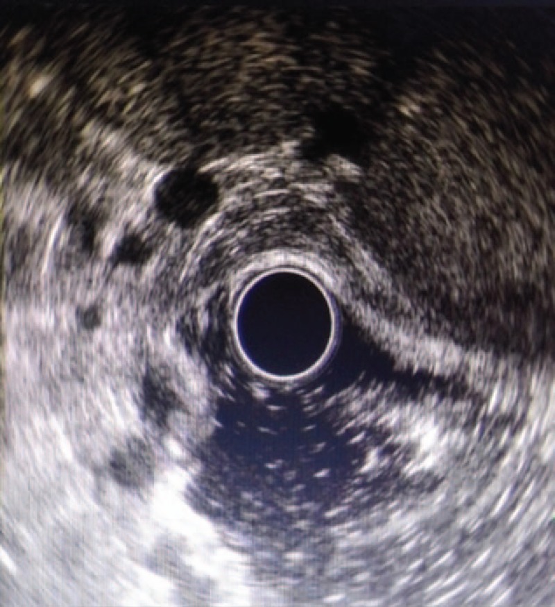
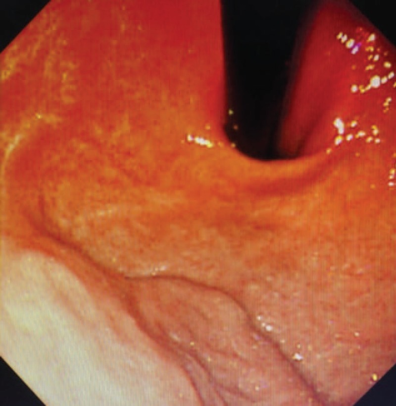
Common endoscopic examination showed no evidence of gastroesophageal varices; EUS revealed distinct varices of the esophageal and gastric veins. Six months after discharge, the patient was rehospitalized because of upper gastrointestinal bleeding. Endoscopic ligation was implemented as well as esophageal varices loop ligature (EVL).

OUTCOMES

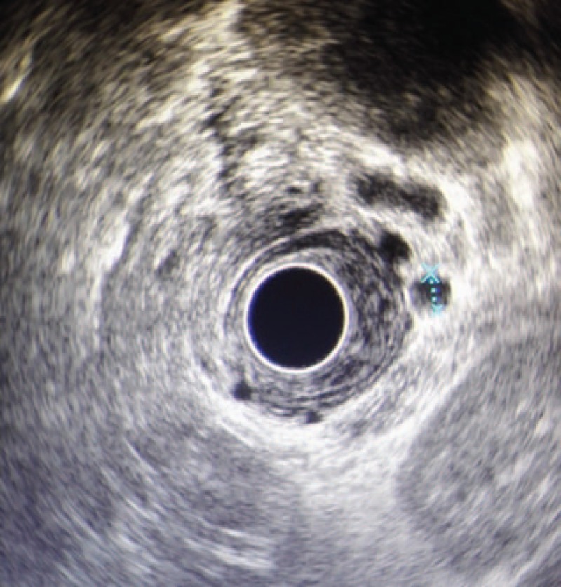
Six months later, EUS showed obvious collateral and perforator veins.

LESSONS

We should strongly recommend that patients with liver cirrhosis undergo EUS in addition to a routine endoscopic examination. EUS can play an important role in evaluating the risk for bleeding in PHT and can be used to assess the efficacy of EVL.

摘要

理论依据

食管和胃静脉曲张破裂出血是肝硬化的严重并发症。既往研究表明,内镜超声检查(EUS)可用于预测肝硬化患者早期食管静脉曲张出血。

患者情况

我们报告一例46岁男性乙肝肝硬化患者(CTP评分5分;Child-Pugh分级为A级),因食欲减退持续1周入院。

诊断

最初诊断为失代偿期乙肝肝硬化;腹部计算机断层扫描(CT)显示肝硬化和门静脉高压(PHT)。

干预措施

普通内镜检查未发现食管胃静脉曲张;EUS显示食管和胃静脉有明显曲张。出院6个月后,患者因上消化道出血再次住院。实施了内镜下套扎术及食管静脉曲张套扎术(EVL)。

结果

6个月后,EUS显示有明显的侧支静脉和穿支静脉。

经验教训

我们强烈建议肝硬化患者除进行常规内镜检查外,还应接受EUS检查。EUS在评估PHT出血风险方面可发挥重要作用,并可用于评估EVL的疗效。

https://cdn.ncbi.nlm.nih.gov/pmc/blobs/b7d8/5413264/c5e527d4462c/medi-96-e6749-g004.jpg
https://cdn.ncbi.nlm.nih.gov/pmc/blobs/b7d8/5413264/c6dc75e31b57/medi-96-e6749-g001.jpg
https://cdn.ncbi.nlm.nih.gov/pmc/blobs/b7d8/5413264/045ec86c5895/medi-96-e6749-g002.jpg
https://cdn.ncbi.nlm.nih.gov/pmc/blobs/b7d8/5413264/c85024e22f33/medi-96-e6749-g003.jpg
https://cdn.ncbi.nlm.nih.gov/pmc/blobs/b7d8/5413264/c5e527d4462c/medi-96-e6749-g004.jpg

相似文献

1
Endoscopic ultrasonography predicts early esophageal variceal bleeding in liver cirrhosis: A case report.内镜超声检查预测肝硬化患者早期食管静脉曲张出血:一例报告。
Medicine (Baltimore). 2017 Apr;96(17):e6749. doi: 10.1097/MD.0000000000006749.
2
The endoscopic ultrasound probe findings in prediction of esophageal variceal recurrence after endoscopic variceal eradication therapies in cirrhotic patients: a cohort prospective study.内镜超声探头检查结果对肝硬化患者内镜下静脉曲张根除治疗后食管静脉曲张复发的预测价值:一项队列前瞻性研究
BMC Gastroenterol. 2019 Feb 19;19(1):32. doi: 10.1186/s12876-019-0943-y.
3
Endoscopic ultrasonographic signs of portal hypertension in cirrhosis.肝硬化门静脉高压的内镜超声征象
Gastrointest Endosc. 1996 Sep;44(3):257-61. doi: 10.1016/s0016-5107(96)70161-x.
4
Utility of endoscopic ultrasound in patients with portal hypertension.内镜超声在门静脉高压患者中的应用价值。
World J Gastroenterol. 2014 Oct 21;20(39):14230-6. doi: 10.3748/wjg.v20.i39.14230.
5
Evaluation and Management of Esophageal and Gastric Varices in Patients with Cirrhosis.肝硬化患者食管胃静脉曲张的评估与管理。
Clin Liver Dis. 2020 Aug;24(3):335-350. doi: 10.1016/j.cld.2020.04.011. Epub 2020 May 31.
6
The role of endoscopic ultrasound for portal hypertension in liver cirrhosis.内镜超声在肝硬化门静脉高压症中的作用。
J Med Ultrason (2001). 2022 Jul;49(3):359-370. doi: 10.1007/s10396-021-01165-4. Epub 2021 Nov 19.
7
Transient Elastography Identifies the Risk of Esophageal Varices and Bleeding in Patients With Hepatitis B Virus-Related Liver Cirrhosis.瞬时弹性成像可识别乙型肝炎病毒相关性肝硬化患者食管静脉曲张及出血风险。
Ultrasound Q. 2018 Sep;34(3):141-147. doi: 10.1097/RUQ.0000000000000373.
8
A modified percutaneous transhepatic variceal embolization with 2-octyl cyanoacrylate versus endoscopic ligation in esophageal variceal bleeding management: randomized controlled trial.使用2-氰基丙烯酸辛酯改良经皮经肝曲张静脉栓塞术与内镜下套扎术治疗食管静脉曲张出血的随机对照试验
Dig Dis Sci. 2008 Aug;53(8):2258-67. doi: 10.1007/s10620-007-0106-9. Epub 2007 Nov 21.
9
Risk Factors for Esophageal Collateral Veins in Cirrhosis with and without Previous Endoscopic Esophageal Variceal Therapy.肝硬化伴或不伴既往内镜食管静脉曲张治疗的食管侧支静脉的危险因素。
Can J Gastroenterol Hepatol. 2022 Jan 4;2022:6666791. doi: 10.1155/2022/6666791. eCollection 2022.
10
Color Doppler ultrasonography is reliable in assessing the risk of esophageal variceal bleeding in patients with liver cirrhosis.彩色多普勒超声检查在评估肝硬化患者食管静脉曲张出血风险方面是可靠的。
Wien Klin Wochenschr. 2005 Oct;117(19-20):711-7. doi: 10.1007/s00508-005-0424-x.

引用本文的文献

1
Correlation between serum liver fibrosis markers and early gastroesophageal varices among patients with compensated liver cirrhosis: a cross-sectional analysis.血清肝纤维化标志物与代偿性肝硬化患者早期胃食管静脉曲张的相关性:一项横断面分析。
BMC Gastroenterol. 2022 Dec 12;22(1):515. doi: 10.1186/s12876-022-02546-w.
2
The Role of Endoscopic Ultrasound for Esophageal Varices.内镜超声在食管静脉曲张中的作用。
Diagnostics (Basel). 2020 Nov 25;10(12):1007. doi: 10.3390/diagnostics10121007.
3
The role of EUS in diagnosis and treatment of liver disorders.

本文引用的文献

1
Utility of endoscopic ultrasound in the diagnosis and management of esophagogastric varices.内镜超声在食管胃静脉曲张诊断和治疗中的应用。
Endosc Ultrasound. 2016 Jul-Aug;5(4):218-24. doi: 10.4103/2303-9027.187840.
2
The role of collateral veins detected by endosonography in predicting the recurrence of esophageal varices after endoscopic treatment: a systematic review.内镜超声检测到的侧支静脉在预测内镜治疗后食管静脉曲张复发中的作用:一项系统评价
Hepatol Int. 2014 Jul;8(3):339-51. doi: 10.1007/s12072-014-9547-3. Epub 2014 Jun 15.
3
Expanding consensus in portal hypertension: Report of the Baveno VI Consensus Workshop: Stratifying risk and individualizing care for portal hypertension.
超声内镜在肝脏疾病诊断与治疗中的作用。
Endosc Int Open. 2019 Oct;7(10):E1262-E1275. doi: 10.1055/a-0958-2183. Epub 2019 Oct 1.
4
Role of combined propofol and sufentanil anesthesia in endoscopic injection sclerotherapy for esophageal varices.丙泊酚联合舒芬太尼麻醉在食管胃底静脉曲张内镜下注射硬化治疗中的作用。
World J Gastroenterol. 2017 Nov 28;23(44):7875-7880. doi: 10.3748/wjg.v23.i44.7875.
门静脉高压领域共识的拓展:巴韦诺VI共识研讨会报告:门静脉高压风险分层与个体化治疗
J Hepatol. 2015 Sep;63(3):743-52. doi: 10.1016/j.jhep.2015.05.022. Epub 2015 Jun 3.
4
Endoscopic Ultrasound (EUS) for Esophageal and Gastric Varices: How Can it Improve the Outcomes and Reduce Complications of Glue Injection.用于食管和胃静脉曲张的内镜超声检查(EUS):它如何改善组织胶注射的效果并减少并发症
J Clin Exp Hepatol. 2012 Mar;2(1):70-4. doi: 10.1016/S0973-6883(12)60083-8. Epub 2012 Apr 12.
5
Endoscopic ultrasound in the diagnosis and treatment of upper digestive bleeding: a useful tool.内镜超声在上消化道出血的诊断和治疗中的应用:一种有用的工具。
J Gastrointestin Liver Dis. 2013 Dec;22(4):465-9.
6
Impact of ethnicity in upper gastrointestinal hemorrhage.种族因素对上消化道出血的影响。
J Clin Gastroenterol. 2014 Apr;48(4):343-50. doi: 10.1097/MCG.0000000000000025.
7
Esophageal variceal ligation in the secondary prevention of variceal bleeding: Result of long term follow-up.食管静脉曲张结扎术用于静脉曲张出血的二级预防:长期随访结果
Pan Afr Med J. 2013 May 3;15:3. doi: 10.11604/pamj.2013.15.3.2098. Print 2013.
8
Mild collateral varices and a fundic plexus without perforating veins on EUS predict endoscopic non-recurrence of esophageal varices after EVL.轻度侧支静脉曲张以及超声内镜检查显示胃底静脉丛无穿支静脉,提示内镜下套扎术后食管静脉曲张不会复发。
Hepatogastroenterology. 2011 May-Jun;58(107-108):798-801.
9
Endoscopic ultrasound (EUS)-guided coil embolization therapy in gastric varices.内镜超声(EUS)引导下胃静脉曲张的线圈栓塞治疗
Endoscopy. 2010;42 Suppl 2:E35-6. doi: 10.1055/s-0029-1215261. Epub 2010 Jan 13.
10
Endoscopic ultrasonographic evaluation of hemodynamics related to variceal relapse in esophageal variceal patients.内镜超声评价与食管静脉曲张患者静脉曲张再发相关的血流动力学。
Hepatol Res. 2009 Feb;39(2):126-33. doi: 10.1111/j.1872-034X.2008.00415.x.