Maadawi Zeinab M El
Department of Histology and Cell Biology, Faculty of Medicine, Cairo University, Cairo, Egypt.
Int J Stem Cells. 2017 May 30;10(1):60-68. doi: 10.15283/ijsc16055.
Neuroinflammation is involved in the pathogenesis of neurodegenerative disorders. Conditioned medium (CM) derived from bone marrow mesenchymal stem cells (MSCs) revealed substantial benefits due to its rich content of trophic factors. Salidroside (Sal), extracted from Rhodiola rosea, is known for its anti-inflammatory and neuroprotective effects. This study was designed to investigate the effect of Sal pretreated CM (CM-Sal) derived from bone marrow MSCs in lipopolysaccharide (LPS) induced neuroinflammation.
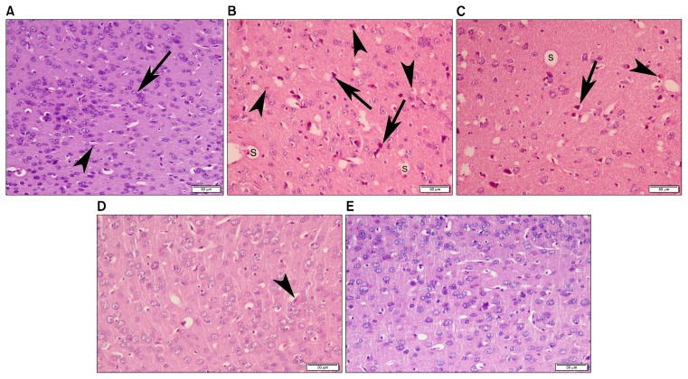
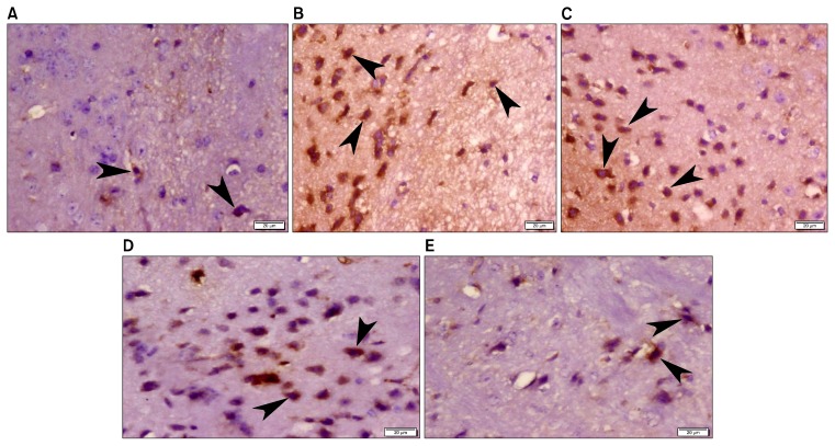
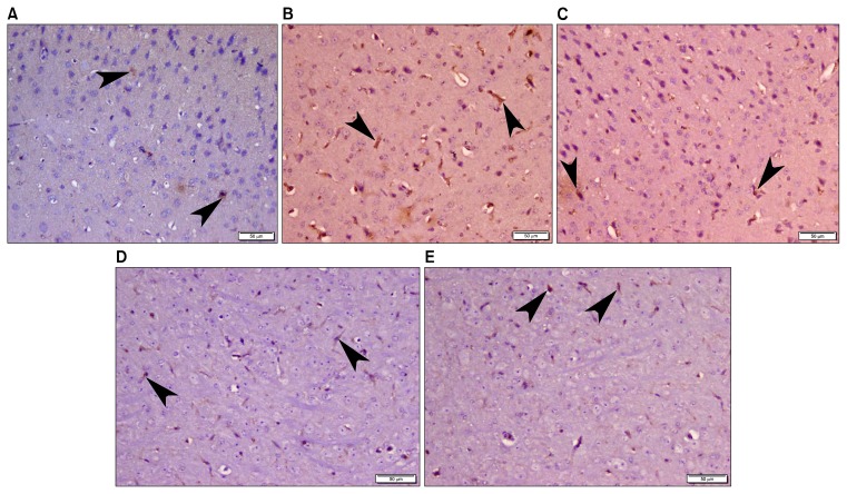
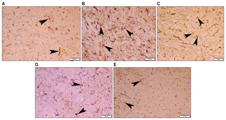
Fifty adult male mice were equally divided into 5 groups: Group I (Normal Control), Group II (LPS): single 0.8 mg/kg LPS intraperitoneally; Group III (LPS-DMEM), Group IV (LPS-CM) and Group V (LPS-CM-Sal): LPS was injected as group II followed, 24 hours later, by intranasal injection of 50 l of filtered serum- free Dulbecco's Modified Eagle's medium (DMEM), CM or CM-Sal, respectively, twice daily for 4 days. Animals were sacrificed at day 6 and paraffin cerebral sections were subjected to Hematoxylin and Eosin staining and immunohistochemistry with caspase 3 (apoptosis), glial fibrillary acidic protein GFAP (astrocytes) and CD68 (active microglia) followed by quantitative morphometric study.
Examination of LPS and LPS-DMEM groups revealed neuronal apoptosis with reactive astrogliosis and increased active microglia. LPS-CM and LPS-CM-Sal groups showed less apoptosis, less astrocytes and less active microglia. The regression in neuroinflammation was more evident in LPS-CM-Sal group and the difference was statistically significant compared to other groups.
CM-Sal derived from MSCs culture elicited significant histopathological improvement in LPS induced neuroinflammation which could be used as new therapeutic modality.
神经炎症参与神经退行性疾病的发病机制。骨髓间充质干细胞(MSCs)分泌的条件培养基(CM)因其富含营养因子而显示出显著益处。红景天苷(Sal)从红景天中提取,以其抗炎和神经保护作用而闻名。本研究旨在探讨骨髓间充质干细胞来源的经红景天苷预处理的条件培养基(CM-Sal)在脂多糖(LPS)诱导的神经炎症中的作用。
50只成年雄性小鼠平均分为5组:第一组(正常对照组);第二组(LPS组):腹腔注射单次0.8mg/kg LPS;第三组(LPS-DMEM组);第四组(LPS-CM组)和第五组(LPS-CM-Sal组):如第二组那样注射LPS,24小时后,分别经鼻内注射50μl过滤后的无血清杜氏改良 Eagle 培养基(DMEM)、CM或CM-Sal,每天两次,共4天。在第6天处死动物,对石蜡脑切片进行苏木精和伊红染色以及用半胱天冬酶3(凋亡)、胶质纤维酸性蛋白GFAP(星形胶质细胞)和CD68(活化小胶质细胞)进行免疫组织化学染色,随后进行定量形态学研究。
LPS组和LPS-DMEM组检查显示神经元凋亡伴有反应性星形胶质细胞增生和活化小胶质细胞增加。LPS-CM组和LPS-CM-Sal组显示凋亡较少、星形胶质细胞较少和活化小胶质细胞较少。神经炎症的消退在LPS-CM-Sal组中更明显,与其他组相比差异有统计学意义。
骨髓间充质干细胞培养产生的CM-Sal在LPS诱导的神经炎症中引起显著的组织病理学改善,可作为一种新的治疗方式。