Hertig Vanessa, Tardif Kim, Meus Marc Andre, Duquette Natacha, Villeneuve Louis, Toussaint Fanny, Ledoux Jonathan, Calderone Angelino
Research Center, Montreal Heart Institute and Université de Montréal, Montréal, Québec, Canada.
Department of Pharmacology & Physiology, Université de Montréal, Québec, Montréal, Canada.
PLoS One. 2017 Apr 27;12(4):e0176147. doi: 10.1371/journal.pone.0176147. eCollection 2017.
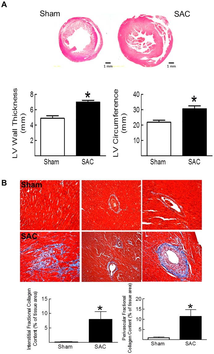
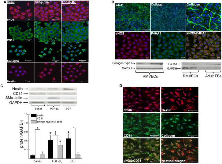
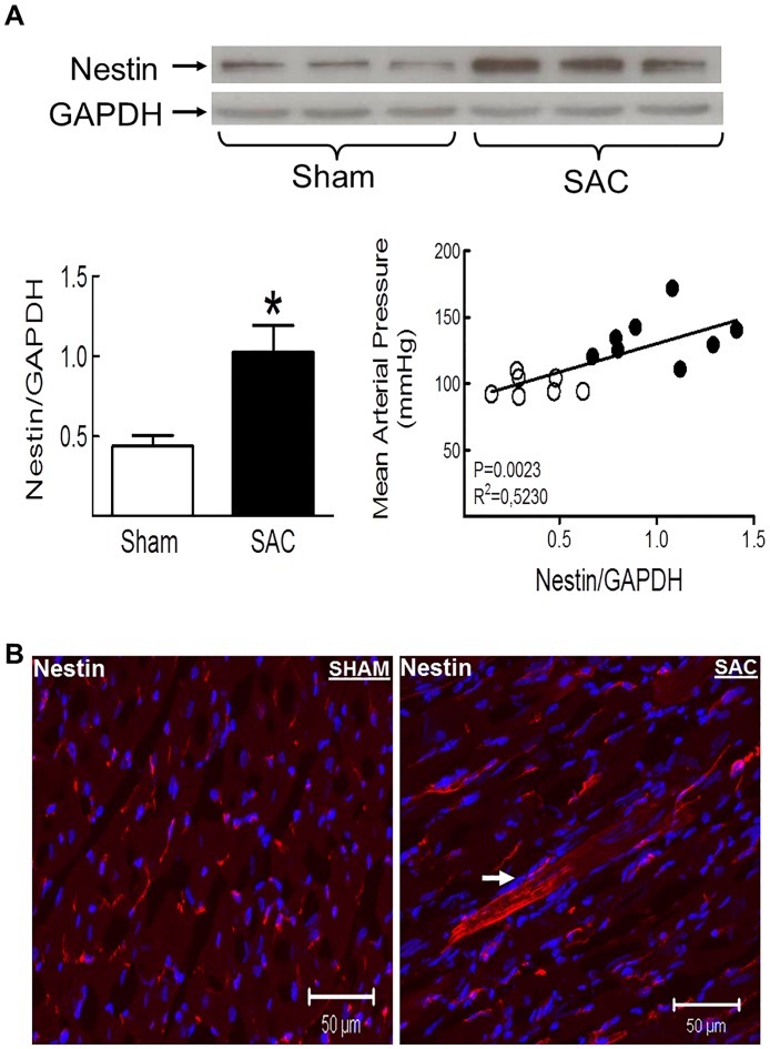
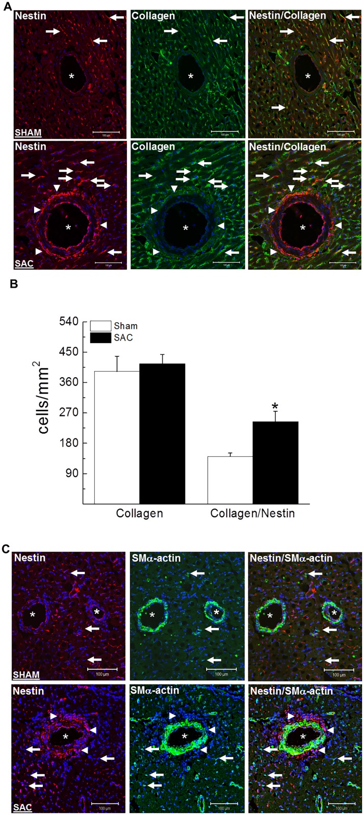
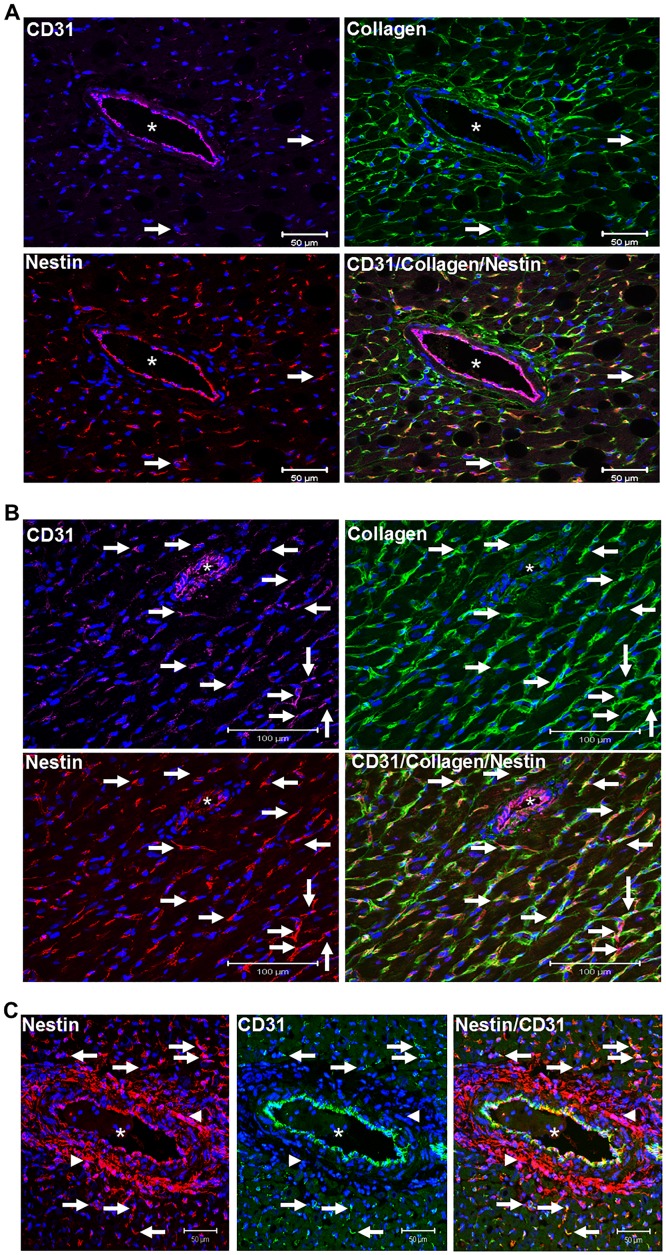
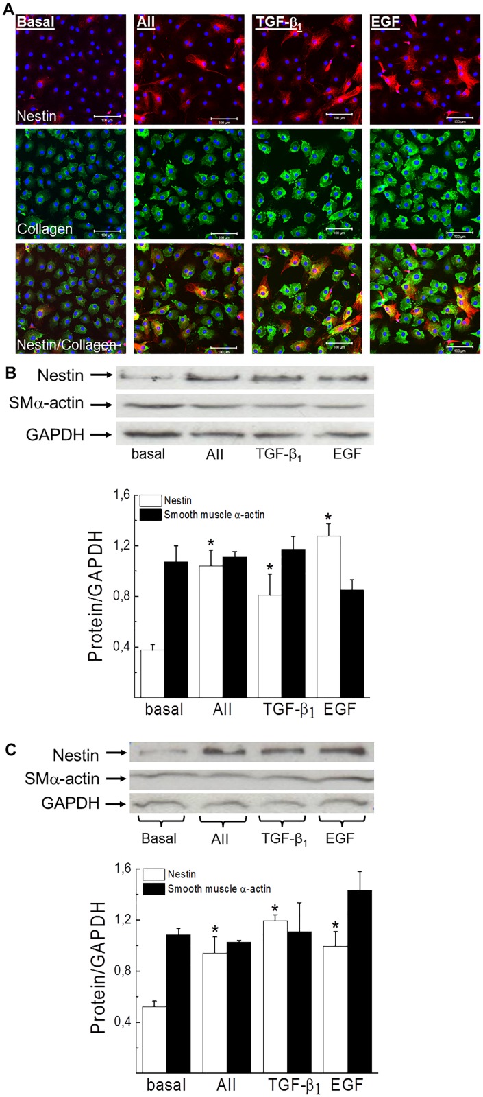
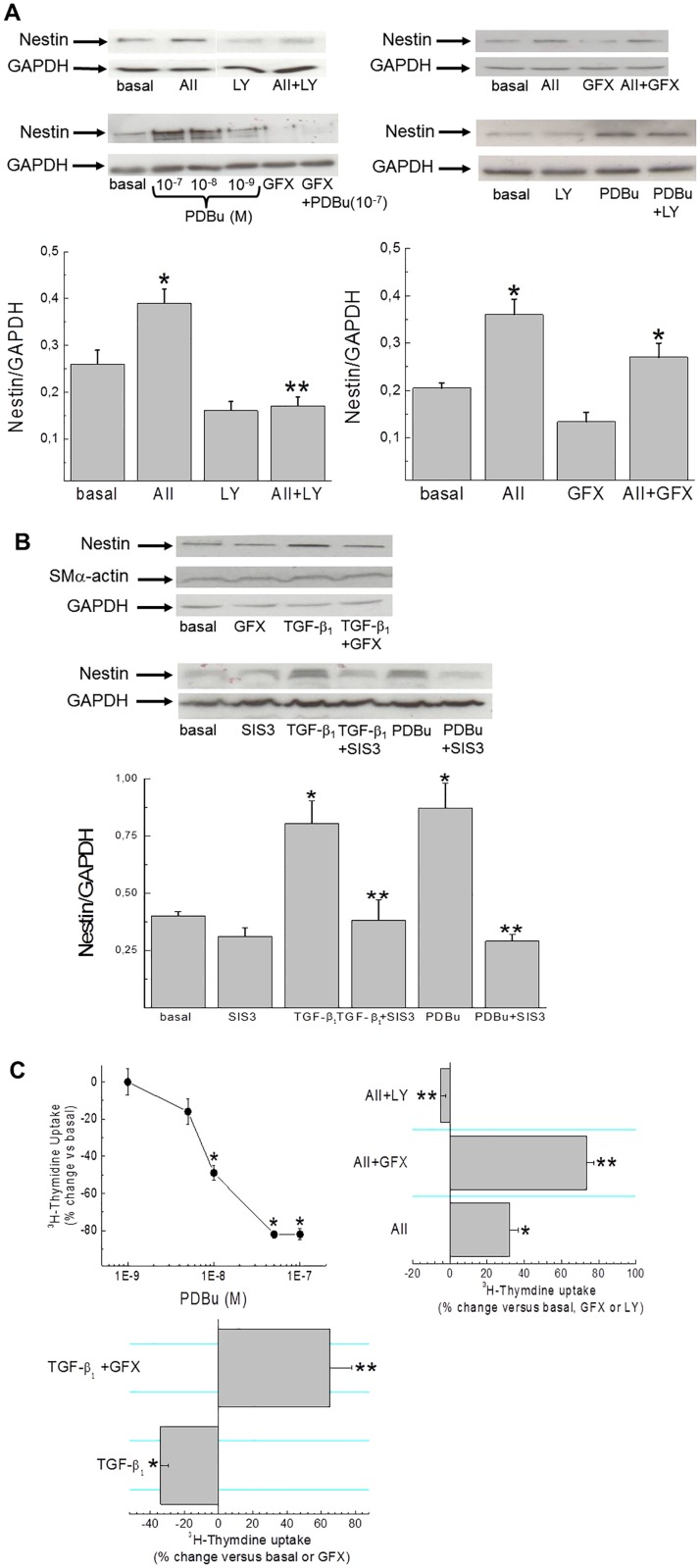
Renal and lung fibrosis was characterized by the accumulation of collagen-immunoreactive mesenchymal cells expressing the intermediate filament protein nestin. The present study tested the hypothesis that nestin expression was increased in the hypertrophied/fibrotic left ventricle of suprarenal abdominal aorta constricted adult male Sprague-Dawley rats and induced in ventricular fibroblasts by pro-fibrotic peptide growth factors. Nestin protein levels were upregulated in the pressure-overloaded left ventricle and expression positively correlated with the rise of mean arterial pressure. In sham and pressure-overloaded hearts, nestin immunoreactivity was detected in collagen type I(+)-and CD31(+)-cells identified in the interstitium and perivascular region whereas staining was absent in smooth muscle α-actin(+)-cells. A significantly greater number of collagen type I(+)-cells co-expressing nestin was identified in the left ventricle of pressure-overloaded rats. Moreover, an accumulation of nestin(+)-cells lacking collagen, CD31 and smooth muscle α-actin staining was selectively observed at the adventitial region of predominantly large calibre blood vessels in the hypertrophied/fibrotic left ventricle. Angiotensin II and TGF-β1 stimulation of ventricular fibroblasts increased nestin protein levels via phosphatidylinositol 3-kinase- and protein kinase C/SMAD3-dependent pathways, respectively. CD31/eNOS(+)-rat cardiac microvascular endothelial cells synthesized/secreted collagen type I, expressed prolyl 4-hydroxylase and TGF-β1 induced nestin expression. The selective accumulation of adventitial nestin(+)-cells highlighted a novel feature of large vessel remodelling in the pressure-overloaded heart and increased appearance of collagen type I/nestin(+)-cells may reflect an activated phenotype of ventricular fibroblasts. CD31/collagen/nestin(+)-interstitial cells could represent displaced endothelial cells displaying an unmasked mesenchymal phenotype, albeit contribution to the reactive fibrotic response of the pressure-overloaded heart remains unknown.
肾和肺纤维化的特征是表达中间丝蛋白巢蛋白的胶原免疫反应性间充质细胞的积累。本研究检验了以下假设:在成年雄性Sprague-Dawley大鼠肾上腹主动脉缩窄所致肥厚/纤维化左心室中,巢蛋白表达增加,且促纤维化肽生长因子可在心室成纤维细胞中诱导其表达。压力超负荷左心室中巢蛋白水平上调,且表达与平均动脉压升高呈正相关。在假手术和压力超负荷心脏中,在间质和血管周围区域鉴定出的I型胶原(+)和CD31(+)细胞中检测到巢蛋白免疫反应性,而在平滑肌α-肌动蛋白(+)细胞中未检测到染色。在压力超负荷大鼠的左心室中,鉴定出大量共表达巢蛋白的I型胶原(+)细胞。此外,在肥厚/纤维化左心室中,主要在大口径血管的外膜区域选择性观察到缺乏胶原、CD31和平滑肌α-肌动蛋白染色的巢蛋白(+)细胞的积累。血管紧张素II和TGF-β1对心室成纤维细胞的刺激分别通过磷脂酰肌醇3-激酶和蛋白激酶C/SMAD3依赖性途径增加巢蛋白水平。CD31/eNOS(+)大鼠心脏微血管内皮细胞合成/分泌I型胶原,表达脯氨酰4-羟化酶,TGF-β1诱导巢蛋白表达。外膜巢蛋白(+)细胞的选择性积累突出了压力超负荷心脏中大血管重塑的一个新特征,I型胶原/巢蛋白(+)细胞出现增加可能反映了心室成纤维细胞的活化表型。CD31/胶原/巢蛋白(+)间质细胞可能代表显示出未掩盖的间充质表型的移位内皮细胞,尽管其对压力超负荷心脏的反应性纤维化反应的贡献仍不清楚。