Department of Orthodontic Science, Graduate School of Medical and Dental Sciences, Tokyo Medical and Dental University, Tokyo, Japan.
Department of Advanced Clinical Science and Therapeutics, Graduate School of Medicine, The University of Tokyo, Tokyo, Japan.
Int J Oral Sci. 2017 Jun;9(2):80-86. doi: 10.1038/ijos.2017.10. Epub 2017 Apr 28.
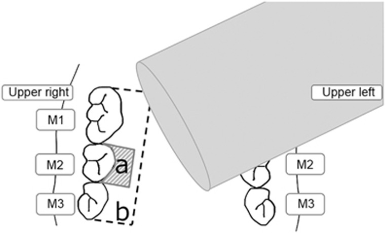
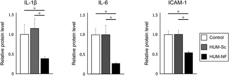
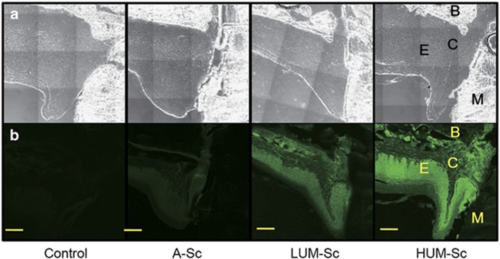
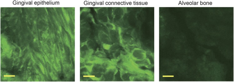
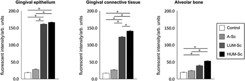
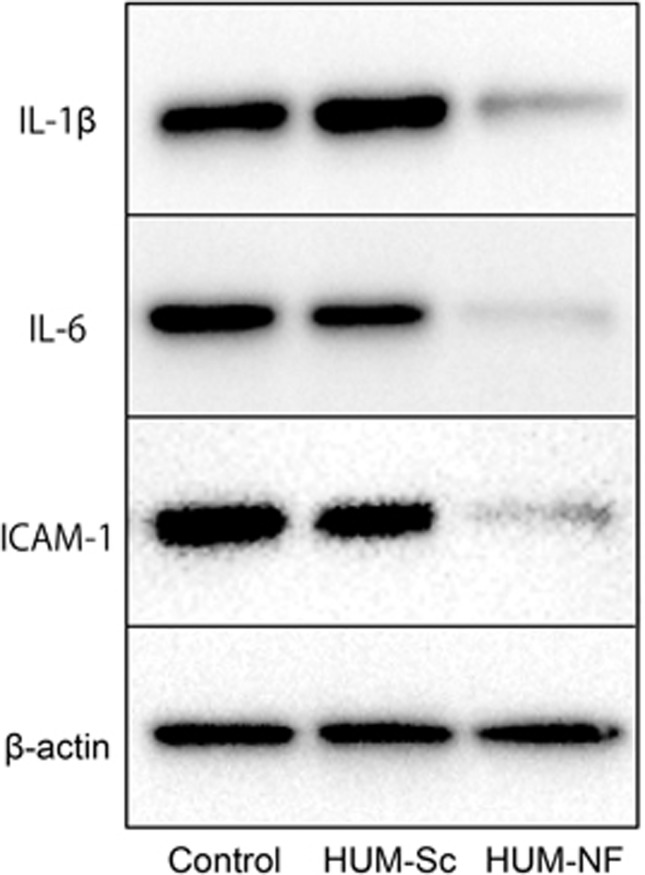
The objective of this study is to investigate the effect of the ultrasound-microbubble technique in nuclear factor kappa B (NF-κB) decoy oligodeoxynucleotide (ODN) transfection in the gingival tissue in mice. The 6-FAM-labeled scrambled decoy ODN with microbubbles was applied to the periodontal tissue in 8-week-old male C57BL/6J mice by ultrasound radiation at low (LUM-Sc) and high (HUM-Sc) intensities to optimize the transfection condition of the ultrasound-microbubble method. Histological inspections were performed two hours after transfection to compare the expression with that in the sham-operated group without ultrasound radiation (A-Sc). Then, an NF-κB decoy was transfected into the periodontal tissue using the high-intensity ultrasound-microbubble (HUM-NF) technique to examine the anti-inflammatory effects of the decoy ODN. Western blot analysis was performed to investigate the expression of interleukin(IL)-1β, IL-6 and intercellular adhesion molecule-1 (ICAM-1) in the gingival tissues in the HUM-Sc, the HUM-NF and control groups. The fluorescence microscopy results showed that the fluorescent intensity in the periodontal tissues in the LUM-Sc and HUM-Sc groups was significantly higher than that in the A-Sc and the control groups. The fluorescent intensity in the HUM-Sc group, especially in the gingival connective tissue, was the highest of all groups. Western blot analysis indicated that the protein expression levels of IL-1β, IL-6 and ICAM-1 in the HUM-NF group were significantly lower than those in the HUM-Sc and the control groups. These findings suggest that the high-intensity ultrasound-microbubble technique is an effective tool for decoy transfection into the periodontal tissue.
本研究旨在探讨超声微泡技术对核因子 kappa B(NF-κB)诱骗寡核苷酸(ODN)转染小鼠牙龈组织的影响。将 6-FAM 标记的乱序诱骗 ODN 与微泡一起应用于 8 周龄雄性 C57BL/6J 小鼠的牙周组织,通过低强度(LUM-Sc)和高强度(HUM-Sc)超声辐射,优化超声微泡法的转染条件。转染后两小时进行组织学检查,以比较与未进行超声辐射的假手术组(A-Sc)的表达情况。然后,采用高强度超声微泡(HUM-NF)技术将 NF-κB 诱骗物转染到牙周组织中,以研究诱骗 ODN 的抗炎作用。采用 Western blot 分析方法研究牙龈组织中白细胞介素(IL)-1β、IL-6 和细胞间黏附分子-1(ICAM-1)的表达。荧光显微镜结果显示,LUM-Sc 和 HUM-Sc 组牙周组织的荧光强度明显高于 A-Sc 和对照组。HUM-Sc 组,尤其是牙龈结缔组织的荧光强度最高。Western blot 分析表明,HUM-NF 组 IL-1β、IL-6 和 ICAM-1 的蛋白表达水平明显低于 HUM-Sc 组和对照组。这些发现表明,高强度超声微泡技术是将诱骗物转染到牙周组织中的有效工具。