Macdonald Ian R, Maxwell Selena P, Reid George A, Cash Meghan K, DeBay Drew R, Darvesh Sultan
Department of Medical Neuroscience, Dalhousie University, Halifax, NS, Canada.
Department of Medicine (Neurology and Geriatric Medicine), Dalhousie University, Halifax, NS, Canada.
J Alzheimers Dis. 2017;58(2):491-505. doi: 10.3233/JAD-170164.
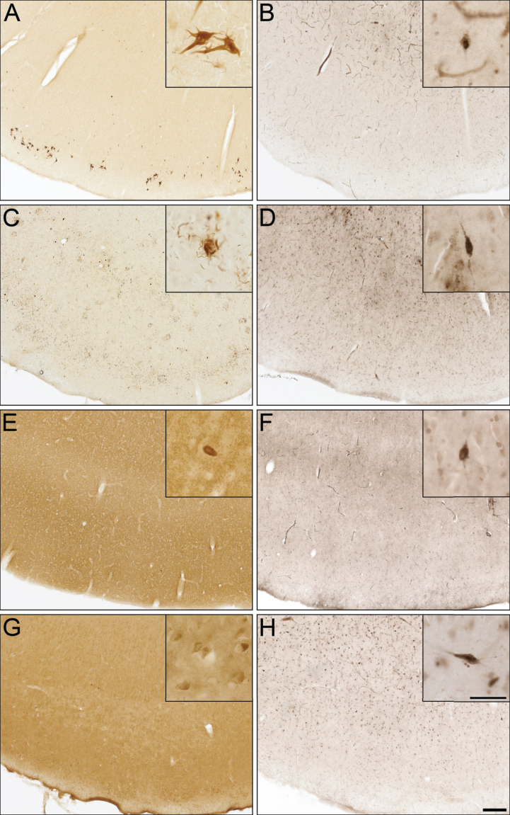
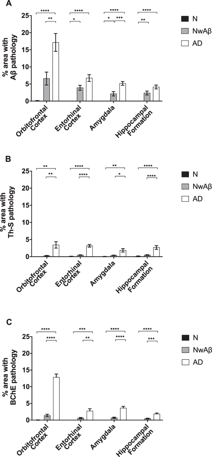
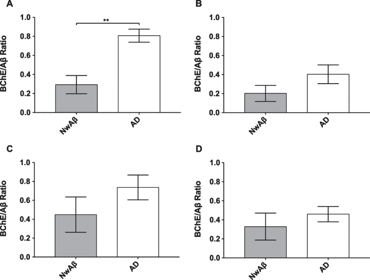
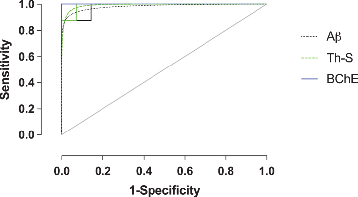
Amyloid-β (Aβ) plaques are a neuropathological hallmark of Alzheimer's disease (AD); however, a significant number of cognitively normal older adults can also have Aβ plaques. Thus, distinguishing AD from cognitively normal individuals with Aβ plaques (NwAβ) based on Aβ plaque detection is challenging. It has been observed that butyrylcholinesterase (BChE) accumulates in plaques preferentially in AD. Thus, detecting BChE-associated plaques has the potential as an improved AD biomarker. We present Aβ, thioflavin-S, and BChE quantification of 26 postmortem brain tissues; AD (n = 8), NwAβ (n = 6), cognitively normal without plaques (n = 8), and other common dementias including corticobasal degeneration, frontotemporal dementia with tau, dementia with Lewy bodies, and vascular dementia. Pathology burden in the orbitofrontal cortex, entorhinal cortex, amygdala, and hippocampal formation was determined and compared. The predictive value of Aβ and BChE quantification was determined, via receiver-operating characteristic plots, to evaluate their AD diagnostic performance using sensitivity, specificity, and area under curve (AUC) metrics. In general, Aβ and BChE-associated pathology were greater in AD, particularly in the orbitofrontal cortex. In this region, the largest increase (9.3-fold) was in BChE-associated pathology, observed between NwAβ and AD, due to the virtual absence of BChE-associated plaques in NwAβ brains. Furthermore, BChE did not associate with pathology of the other dementias. In this sample, BChE-associated pathology provided better diagnostic performance (AUC = 1.0, sensitivity/specificity = 100% /100%) when compared to Aβ (AUC = 0.98, 100% /85.7%). These findings highlight the predictive value of BChE as a biomarker for AD that could facilitate timely disease diagnosis and management.
淀粉样蛋白-β(Aβ)斑块是阿尔茨海默病(AD)的神经病理学标志;然而,相当数量认知正常的老年人也可能有Aβ斑块。因此,基于Aβ斑块检测将AD与有Aβ斑块的认知正常个体(NwAβ)区分开来具有挑战性。据观察,丁酰胆碱酯酶(BChE)优先在AD的斑块中积累。因此,检测与BChE相关的斑块有潜力成为一种改进的AD生物标志物。我们展示了对26个死后脑组织中Aβ、硫黄素-S和BChE的定量分析;包括AD(n = 8)、NwAβ(n = 6)、无斑块的认知正常个体(n = 8),以及其他常见痴呆症,包括皮质基底节变性、tau蛋白相关的额颞叶痴呆、路易体痴呆和血管性痴呆。确定并比较了眶额皮质、内嗅皮质、杏仁核和海马结构中的病理负担。通过受试者工作特征曲线确定Aβ和BChE定量的预测价值,以使用敏感性、特异性和曲线下面积(AUC)指标评估它们的AD诊断性能。总体而言,AD中与Aβ和BChE相关的病理情况更严重,尤其是在眶额皮质。在该区域,NwAβ和AD之间观察到与BChE相关的病理情况增加幅度最大(9.3倍),这是由于NwAβ脑内几乎不存在与BChE相关的斑块。此外,BChE与其他痴呆症的病理情况无关。在这个样本中,与Aβ(AUC = 0.98,100% / 85.7%)相比,与BChE相关的病理情况提供了更好的诊断性能(AUC = 1.0,敏感性/特异性 = 100% / 100%)。这些发现突出了BChE作为AD生物标志物的预测价值,这有助于及时进行疾病诊断和管理。