• 文献检索
  • 文档翻译
  • 深度研究
  • 学术资讯
  • Suppr Zotero 插件Zotero 插件
  • 邀请有礼
  • 套餐&价格
  • 历史记录
应用&插件
Suppr Zotero 插件Zotero 插件浏览器插件Mac 客户端Windows 客户端微信小程序
定价
高级版会员购买积分包购买API积分包
服务
文献检索文档翻译深度研究API 文档MCP 服务
关于我们
关于 Suppr公司介绍联系我们用户协议隐私条款
关注我们

Suppr 超能文献

核心技术专利:CN118964589B侵权必究
粤ICP备2023148730 号-1Suppr @ 2026

文献检索

告别复杂PubMed语法,用中文像聊天一样搜索,搜遍4000万医学文献。AI智能推荐,让科研检索更轻松。

立即免费搜索

文件翻译

保留排版,准确专业,支持PDF/Word/PPT等文件格式,支持 12+语言互译。

免费翻译文档

深度研究

AI帮你快速写综述,25分钟生成高质量综述,智能提取关键信息,辅助科研写作。

立即免费体验

衰老与疾病中衰老细胞的定量鉴定。

Quantitative identification of senescent cells in aging and disease.

作者信息

Biran Anat, Zada Lior, Abou Karam Paula, Vadai Ezra, Roitman Lior, Ovadya Yossi, Porat Ziv, Krizhanovsky Valery

机构信息

Department of Molecular Cell Biology, Weizmann Institute of Science, Rehovot, 76100, Israel.

Flow Cytometry Unit, Biological Services Department, Weizmann Institute of Science, 76100, Rehovot, Israel.

出版信息

Aging Cell. 2017 Aug;16(4):661-671. doi: 10.1111/acel.12592. Epub 2017 Apr 28.

DOI:10.1111/acel.12592
PMID:28455874
原文链接:https://pmc.ncbi.nlm.nih.gov/articles/PMC5506427/
Abstract

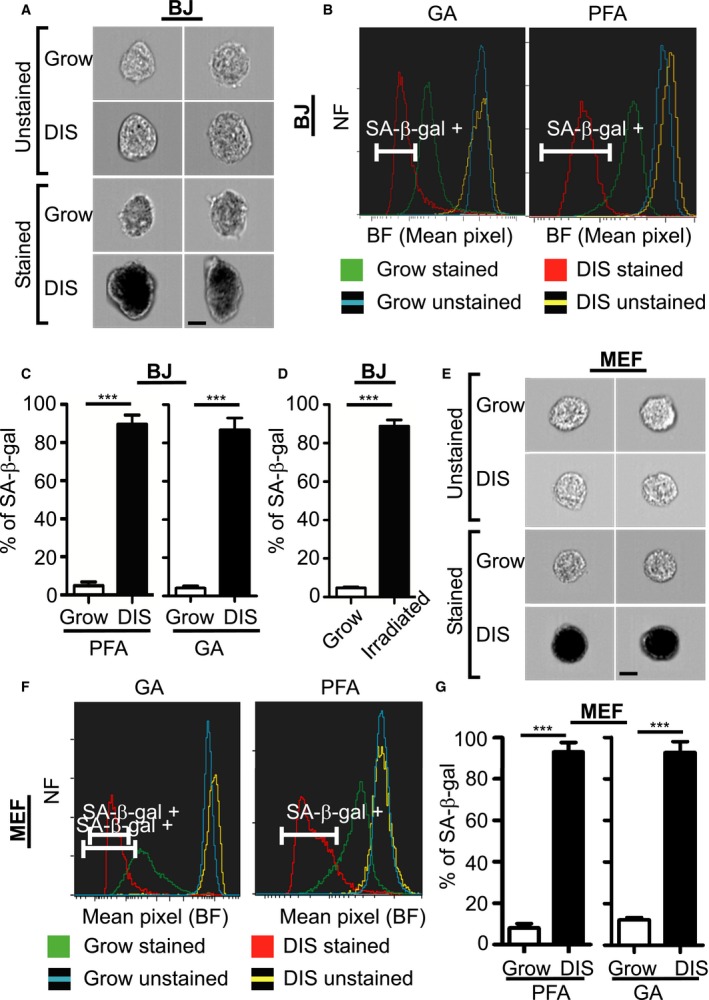
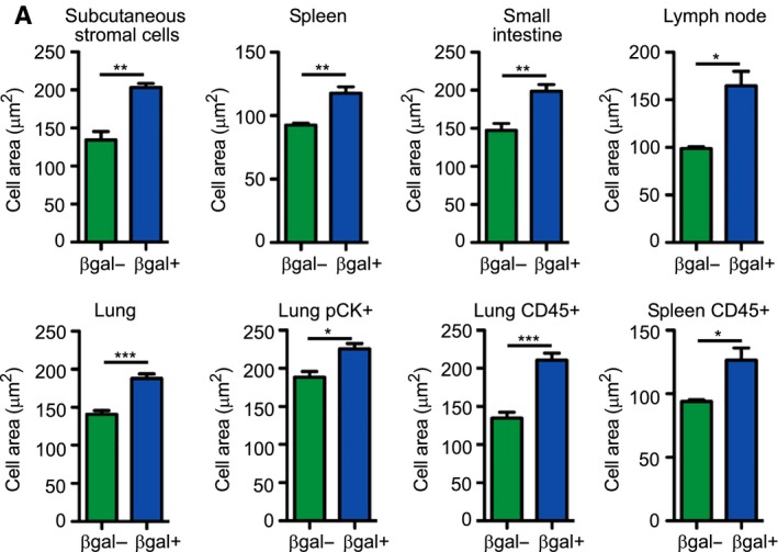
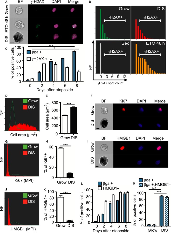
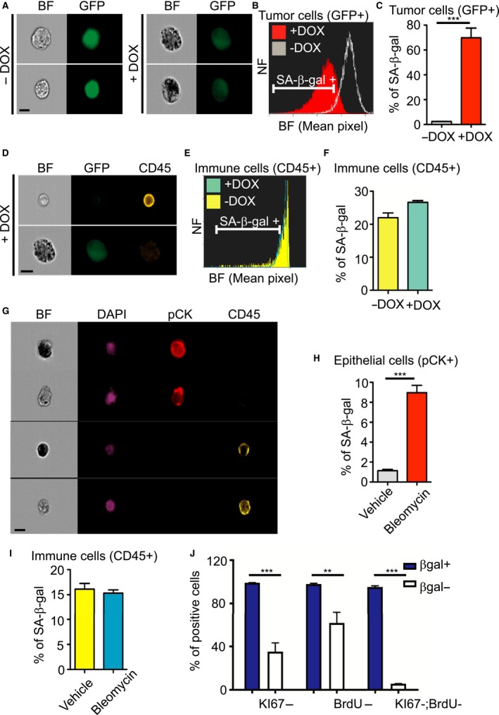
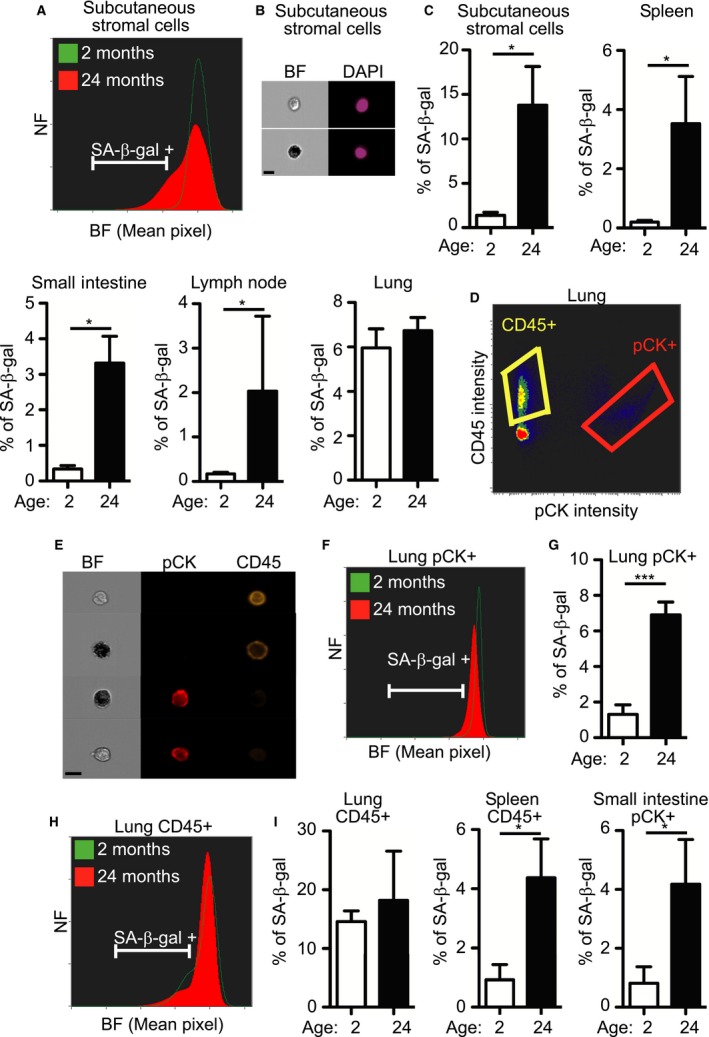
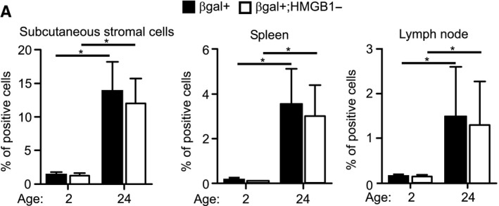
Senescent cells are present in premalignant lesions and sites of tissue damage and accumulate in tissues with age. In vivo identification, quantification and characterization of senescent cells are challenging tasks that limit our understanding of the role of senescent cells in diseases and aging. Here, we present a new way to precisely quantify and identify senescent cells in tissues on a single-cell basis. The method combines a senescence-associated beta-galactosidase assay with staining of molecular markers for cellular senescence and of cellular identity. By utilizing technology that combines flow cytometry with high-content image analysis, we were able to quantify senescent cells in tumors, fibrotic tissues, and tissues of aged mice. Our approach also yielded the finding that senescent cells in tissues of aged mice are larger than nonsenescent cells. Thus, this method provides a basis for quantitative assessment of senescent cells and it offers proof of principle for combination of different markers of senescence. It paves the way for screening of senescent cells for identification of new senescence biomarkers, genes that bypass senescence or senolytic compounds that eliminate senescent cells, thus enabling a deeper understanding of the senescent state in vivo.

摘要

衰老细胞存在于癌前病变和组织损伤部位,并随着年龄的增长在组织中积累。衰老细胞的体内鉴定、定量和表征是具有挑战性的任务,限制了我们对衰老细胞在疾病和衰老中作用的理解。在这里,我们提出了一种在单细胞水平上精确量化和鉴定组织中衰老细胞的新方法。该方法将衰老相关β-半乳糖苷酶检测与细胞衰老分子标记和细胞身份标记的染色相结合。通过利用将流式细胞术与高内涵图像分析相结合的技术,我们能够对肿瘤、纤维化组织和老年小鼠组织中的衰老细胞进行定量。我们的方法还发现老年小鼠组织中的衰老细胞比非衰老细胞大。因此,该方法为衰老细胞的定量评估提供了基础,并为不同衰老标记物的组合提供了原理证明。它为筛选衰老细胞以鉴定新的衰老生物标志物、绕过衰老的基因或消除衰老细胞的衰老溶解化合物铺平了道路,从而能够更深入地了解体内的衰老状态。

https://cdn.ncbi.nlm.nih.gov/pmc/blobs/33f0/5506427/ba2a2aaf655a/ACEL-16-661-g006.jpg
https://cdn.ncbi.nlm.nih.gov/pmc/blobs/33f0/5506427/635b305aad4c/ACEL-16-661-g001.jpg
https://cdn.ncbi.nlm.nih.gov/pmc/blobs/33f0/5506427/56f14cb2de00/ACEL-16-661-g002.jpg
https://cdn.ncbi.nlm.nih.gov/pmc/blobs/33f0/5506427/b1f990c038d2/ACEL-16-661-g003.jpg
https://cdn.ncbi.nlm.nih.gov/pmc/blobs/33f0/5506427/461256ee94c9/ACEL-16-661-g004.jpg
https://cdn.ncbi.nlm.nih.gov/pmc/blobs/33f0/5506427/d24520f39035/ACEL-16-661-g005.jpg
https://cdn.ncbi.nlm.nih.gov/pmc/blobs/33f0/5506427/ba2a2aaf655a/ACEL-16-661-g006.jpg

相似文献

1
Quantitative identification of senescent cells in aging and disease.衰老与疾病中衰老细胞的定量鉴定。
Aging Cell. 2017 Aug;16(4):661-671. doi: 10.1111/acel.12592. Epub 2017 Apr 28.
2
A Multiparametric Assay to Evaluate Senescent Cells.一种评估衰老细胞的多参数检测方法。
Methods Mol Biol. 2019;1896:107-117. doi: 10.1007/978-1-4939-8931-7_11.
3
Quantitative Identification of Senescent Cells in Cancer.癌症中衰老细胞的定量鉴定
Methods Mol Biol. 2019;1884:259-267. doi: 10.1007/978-1-4939-8885-3_18.
4
Analysis of individual cells identifies cell-to-cell variability following induction of cellular senescence.分析单个细胞可鉴定细胞衰老诱导后的细胞间变异性。
Aging Cell. 2017 Oct;16(5):1043-1050. doi: 10.1111/acel.12632. Epub 2017 Jul 11.
5
Multiparameter flow cytometric detection and quantification of senescent cells in vitro.体外多参数流式细胞术检测和定量衰老细胞。
Biogerontology. 2020 Dec;21(6):773-786. doi: 10.1007/s10522-020-09893-9. Epub 2020 Aug 10.
6
Detection of Senescent Cells by Extracellular Markers Using a Flow Cytometry-Based Approach.基于流式细胞术的方法通过细胞外标志物检测衰老细胞
Methods Mol Biol. 2017;1534:147-153. doi: 10.1007/978-1-4939-6670-7_14.
7
Phenotypic and functional differences between senescent and aged murine microglia.衰老和老年小鼠小胶质细胞之间表型和功能的差异。
Neurobiol Aging. 2019 Feb;74:56-69. doi: 10.1016/j.neurobiolaging.2018.10.007. Epub 2018 Oct 12.
8
Overexpression of the novel senescence marker β-galactosidase (GLB1) in prostate cancer predicts reduced PSA recurrence.新型衰老标志物β-半乳糖苷酶(GLB1)在前列腺癌中的过表达预示着前列腺特异性抗原(PSA)复发率降低。
PLoS One. 2015 Apr 15;10(4):e0124366. doi: 10.1371/journal.pone.0124366. eCollection 2015.
9
Loss of lamin B1 is a biomarker to quantify cellular senescence in photoaged skin. lamin B1 的缺失是量化光老化皮肤细胞衰老的生物标志物。
Sci Rep. 2017 Nov 15;7(1):15678. doi: 10.1038/s41598-017-15901-9.
10
Hallmarks of Cellular Senescence.细胞衰老的特征。
Trends Cell Biol. 2018 Jun;28(6):436-453. doi: 10.1016/j.tcb.2018.02.001. Epub 2018 Feb 21.

引用本文的文献

1
An effective system for senescence modulating drug development using quantitative high-content analysis and high-throughput screening.一种利用定量高内涵分析和高通量筛选进行衰老调节药物开发的有效系统。
Commun Biol. 2025 Aug 30;8(1):1316. doi: 10.1038/s42003-025-08758-6.
2
Characterizing primary and secondary senescence in vivo.体内原发性和继发性衰老的特征分析。
Nat Aging. 2025 Jul 11. doi: 10.1038/s43587-025-00917-y.
3
Hematopoietic stem cell size heterogeneity is not linked to changes in stem cell potential of aged HSCs.造血干细胞大小的异质性与衰老造血干细胞的干细胞潜能变化无关。

本文引用的文献

1
Directed elimination of senescent cells by inhibition of BCL-W and BCL-XL.通过抑制BCL-W和BCL-XL定向清除衰老细胞。
Nat Commun. 2016 Apr 6;7:11190. doi: 10.1038/ncomms11190.
2
NKG2D ligands mediate immunosurveillance of senescent cells.NKG2D配体介导衰老细胞的免疫监视。
Aging (Albany NY). 2016 Feb;8(2):328-44. doi: 10.18632/aging.100897.
3
Naturally occurring p16(Ink4a)-positive cells shorten healthy lifespan.天然存在的p16(Ink4a)阳性细胞会缩短健康寿命。
Front Aging. 2025 May 20;6:1596565. doi: 10.3389/fragi.2025.1596565. eCollection 2025.
4
p21-Dependent Senescence Induction by BMP4 Renders Glioblastoma Cells Vulnerable to Senolytics.BMP4通过p21依赖性诱导衰老使胶质母细胞瘤细胞易受衰老溶解剂的影响。
Int J Mol Sci. 2025 Apr 23;26(9):3974. doi: 10.3390/ijms26093974.
5
ABT263 Ameliorates Cellular Senescence, Aβ-Dependent Pathology and Cognitive Decline in Aged APP/PS1 Mice via Regulating PI3K/AKT/GSK-3β Pathways.ABT263通过调节PI3K/AKT/GSK-3β信号通路改善老年APP/PS1小鼠的细胞衰老、Aβ相关病理变化及认知衰退。
Cell Biochem Biophys. 2025 Apr 28. doi: 10.1007/s12013-025-01745-y.
6
An unbiased cell-culture selection yields DNA aptamers as novel senescent cell-specific reagents.无偏差的细胞培养筛选产生DNA适配体,作为新型衰老细胞特异性试剂。
bioRxiv. 2025 Mar 19:2025.03.19.643361. doi: 10.1101/2025.03.19.643361.
7
Targeting senescent hepatocytes for treatment of metabolic dysfunction-associated steatotic liver disease and multi-organ dysfunction.靶向衰老肝细胞治疗代谢功能障碍相关脂肪性肝病和多器官功能障碍。
Nat Commun. 2025 Mar 28;16(1):3038. doi: 10.1038/s41467-025-57616-w.
8
The human-specific noncoding RNA functions at the intersection of sexually dimorphic pathways in inflammation, senescence, and metabolism.人类特有的非编码RNA在炎症、衰老和新陈代谢的性别二态性途径的交叉点发挥作用。
FASEB Bioadv. 2025 Jan 8;7(2):e1479. doi: 10.1096/fba.2024-00101. eCollection 2025 Feb.
9
Aging Lung: Molecular Drivers and Impact on Respiratory Diseases-A Narrative Clinical Review.衰老的肺脏:分子驱动因素及其对呼吸系统疾病的影响——一篇叙述性临床综述
Antioxidants (Basel). 2024 Dec 2;13(12):1480. doi: 10.3390/antiox13121480.
10
Senolytic treatment attenuates immune cell infiltration without improving IAV outcomes in aged mice.衰老细胞清除疗法可减轻老年小鼠的免疫细胞浸润,但不会改善甲型流感病毒感染的结果。
Aging Cell. 2025 Apr;24(4):e14437. doi: 10.1111/acel.14437. Epub 2025 Jan 3.
Nature. 2016 Feb 11;530(7589):184-9. doi: 10.1038/nature16932. Epub 2016 Feb 3.
4
Clearance of senescent cells by ABT263 rejuvenates aged hematopoietic stem cells in mice.ABT263清除衰老细胞可使小鼠体内衰老的造血干细胞恢复活力。
Nat Med. 2016 Jan;22(1):78-83. doi: 10.1038/nm.4010. Epub 2015 Dec 14.
5
Senescent cells communicate via intercellular protein transfer.衰老细胞通过细胞间蛋白质转移进行通讯。
Genes Dev. 2015 Apr 15;29(8):791-802. doi: 10.1101/gad.259341.115. Epub 2015 Apr 8.
6
Physiological and pathological consequences of cellular senescence.细胞衰老的生理和病理后果。
Cell Mol Life Sci. 2014 Nov;71(22):4373-86. doi: 10.1007/s00018-014-1691-3. Epub 2014 Jul 31.
7
Cellular senescence: from physiology to pathology.细胞衰老:从生理学到病理学。
Nat Rev Mol Cell Biol. 2014 Jul;15(7):482-96. doi: 10.1038/nrm3823.
8
Cellular senescence and its effector programs.细胞衰老及其效应程序。
Genes Dev. 2014 Jan 15;28(2):99-114. doi: 10.1101/gad.235184.113.
9
Senescence-associated secretory phenotype in a mouse model of bleomycin-induced lung injury.博来霉素诱导的小鼠肺损伤模型中的衰老相关分泌表型
Exp Toxicol Pathol. 2013 Nov;65(7-8):1053-62. doi: 10.1016/j.etp.2013.04.001. Epub 2013 May 18.
10
p53-dependent release of Alarmin HMGB1 is a central mediator of senescent phenotypes.p53 依赖性 HMGB1 警报素的释放是衰老表型的核心介质。
J Cell Biol. 2013 May 13;201(4):613-29. doi: 10.1083/jcb.201206006. Epub 2013 May 6.